
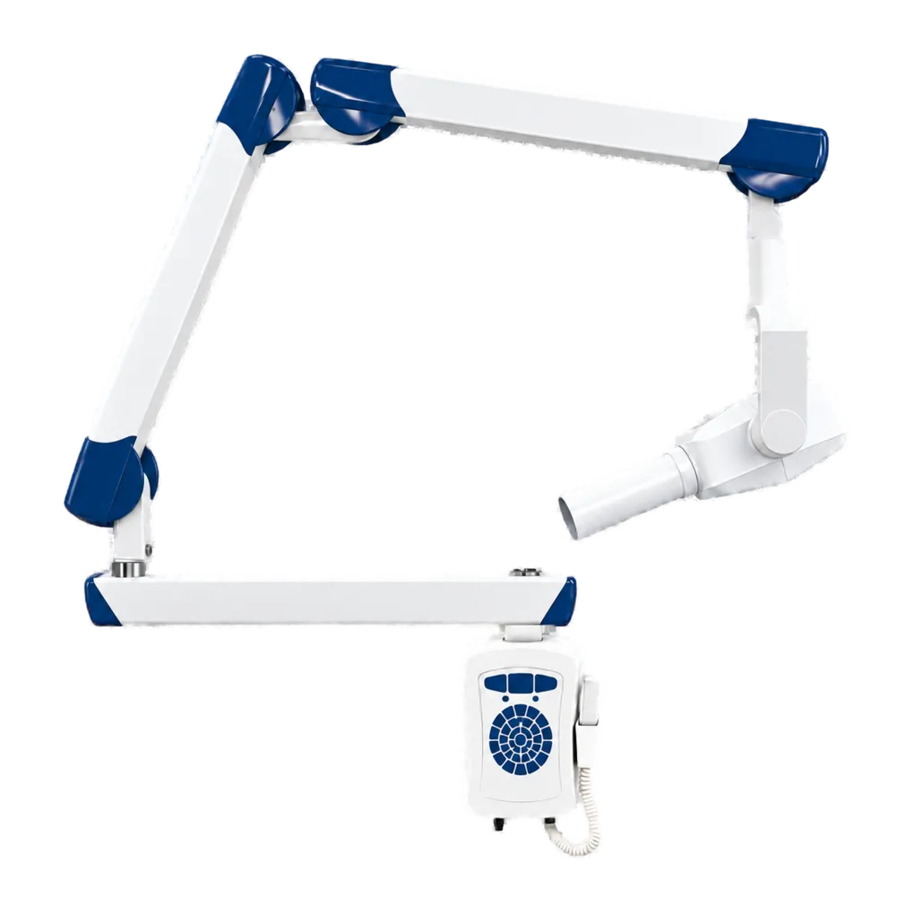
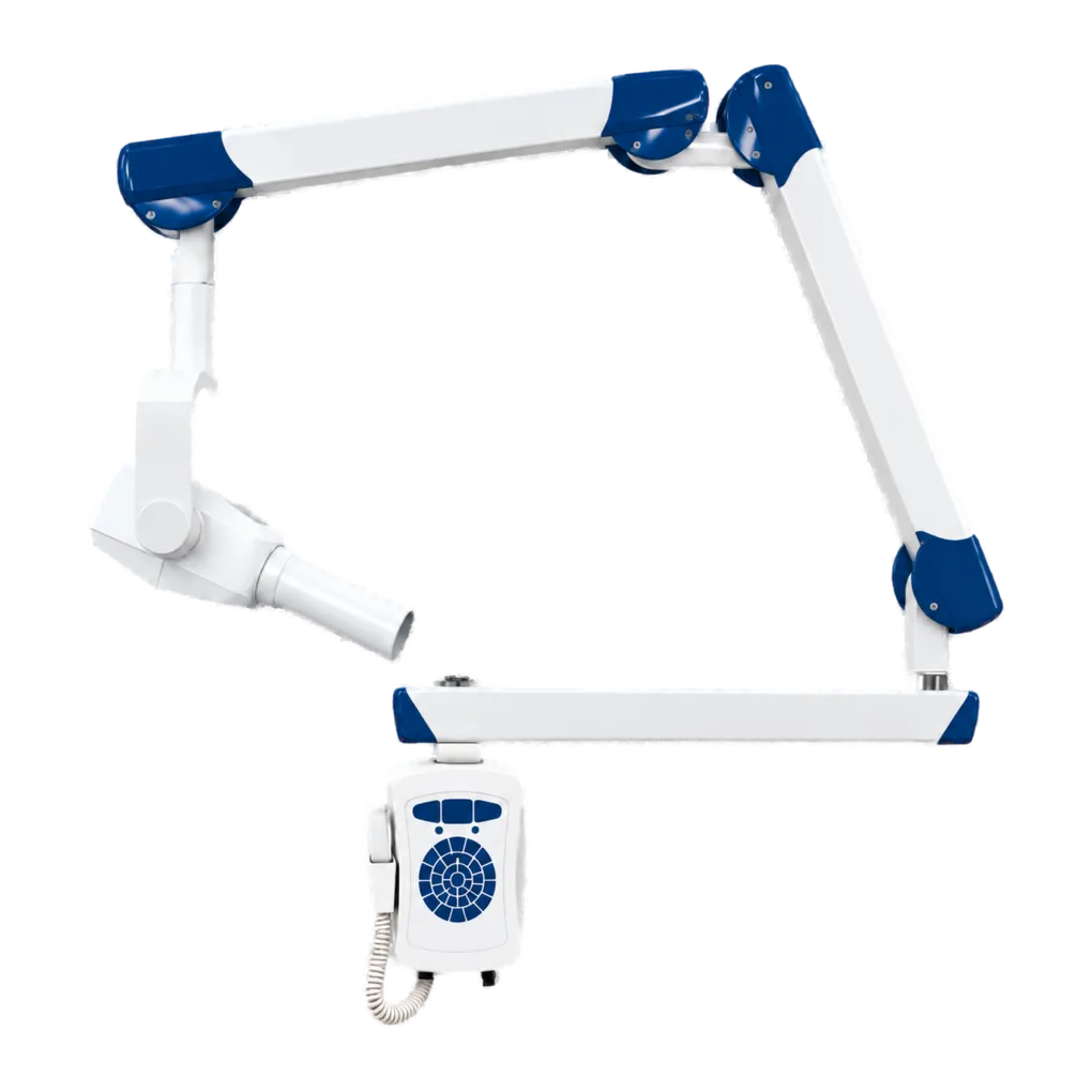
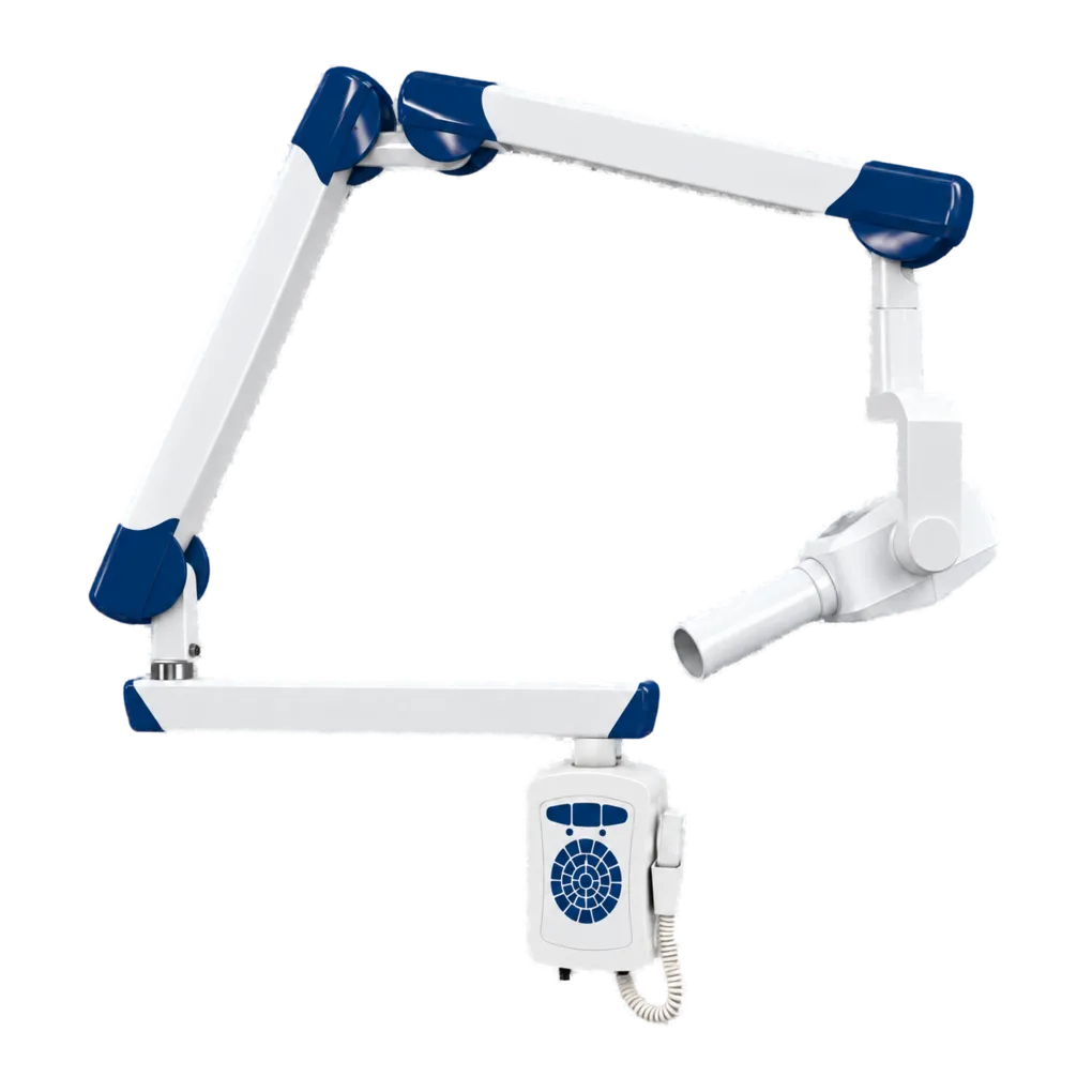
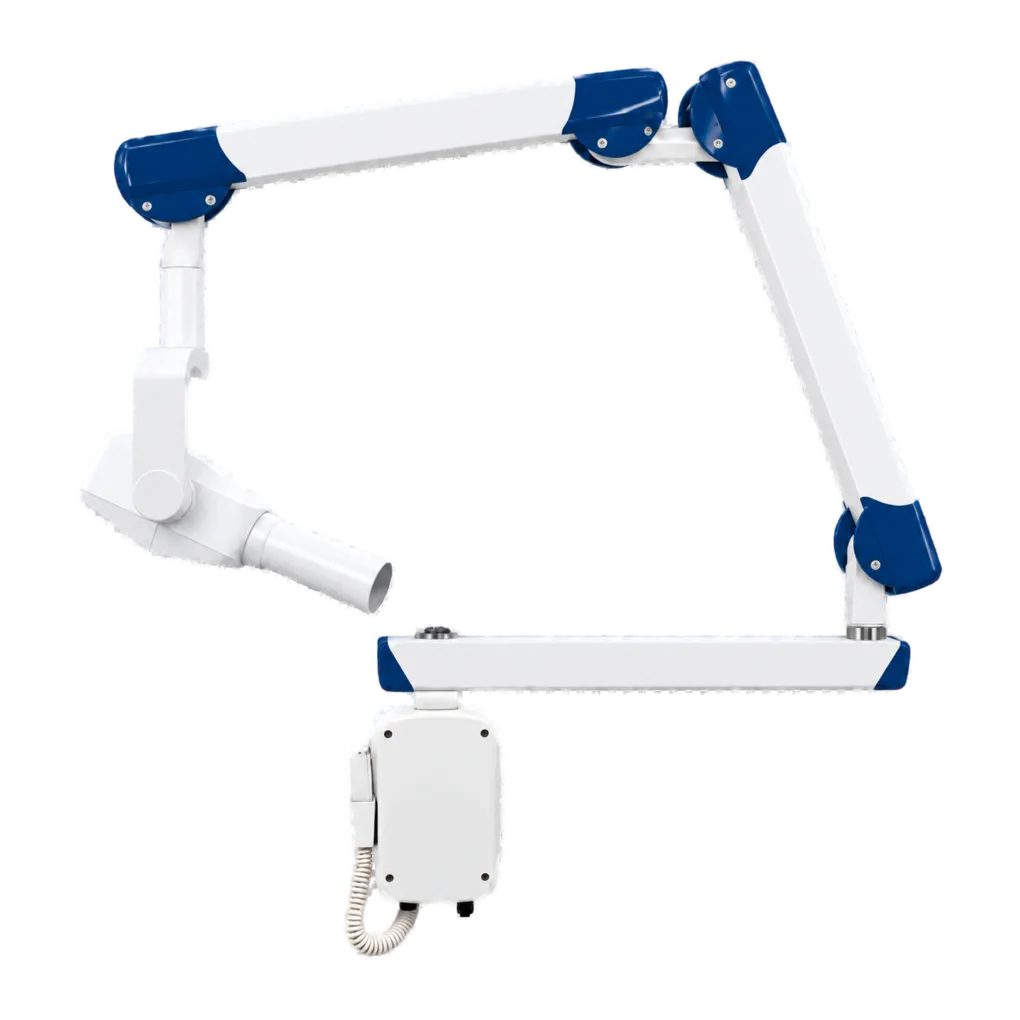
Intraoral radiography carries the diagnostic load in UK general dentistry, and a wall-mounted unit is the space-efficient way to install it in a surgery. The SJD-C09D supports bitewing caries detection, periapical assessment of pathology, endodontic working-length and obturation verification, alveolar bone-level review, and root morphology assessment ahead of extraction. The manufacturer specifically identifies root canal work among its intended uses.
Operation at 70 kV shapes the images. A higher-energy beam produces a longer greyscale, which suits views where crestal bone, root surface and restorative material have to be read together — periodontal assessment and restorative margin review both benefit — and it reaches a given receptor density in a shorter exposure than a lower kilovoltage would.
The same kilovoltage sets the installation bar, and this is the point to resolve before committing. Equipment above 60 kV attracts the longer minimum focus-to-skin distance, and no cone or distance figure is published. That is a specification question rather than a clinical one, but it determines whether the unit can be used as supplied.
The wall-mounted format suits surgeries where floor space is limited, folding back between patients and extending over the chair when needed. The trade-off is that the installation is fixed: the arm's reach has to cover every chair position it will serve, which is worth checking against a scale plan.
Remote exposure triggering is the format's real dose advantage, letting the operator stand outside the controlled area for every exposure. Guidance from the College of General Dentistry and UKHSA Dental X-ray Protection Services remains the reference point UK practices work to, alongside HSE enforcement of IRR17.