
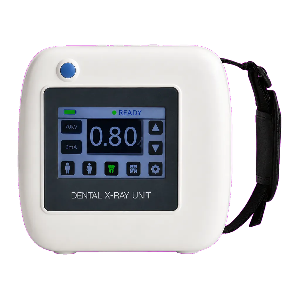
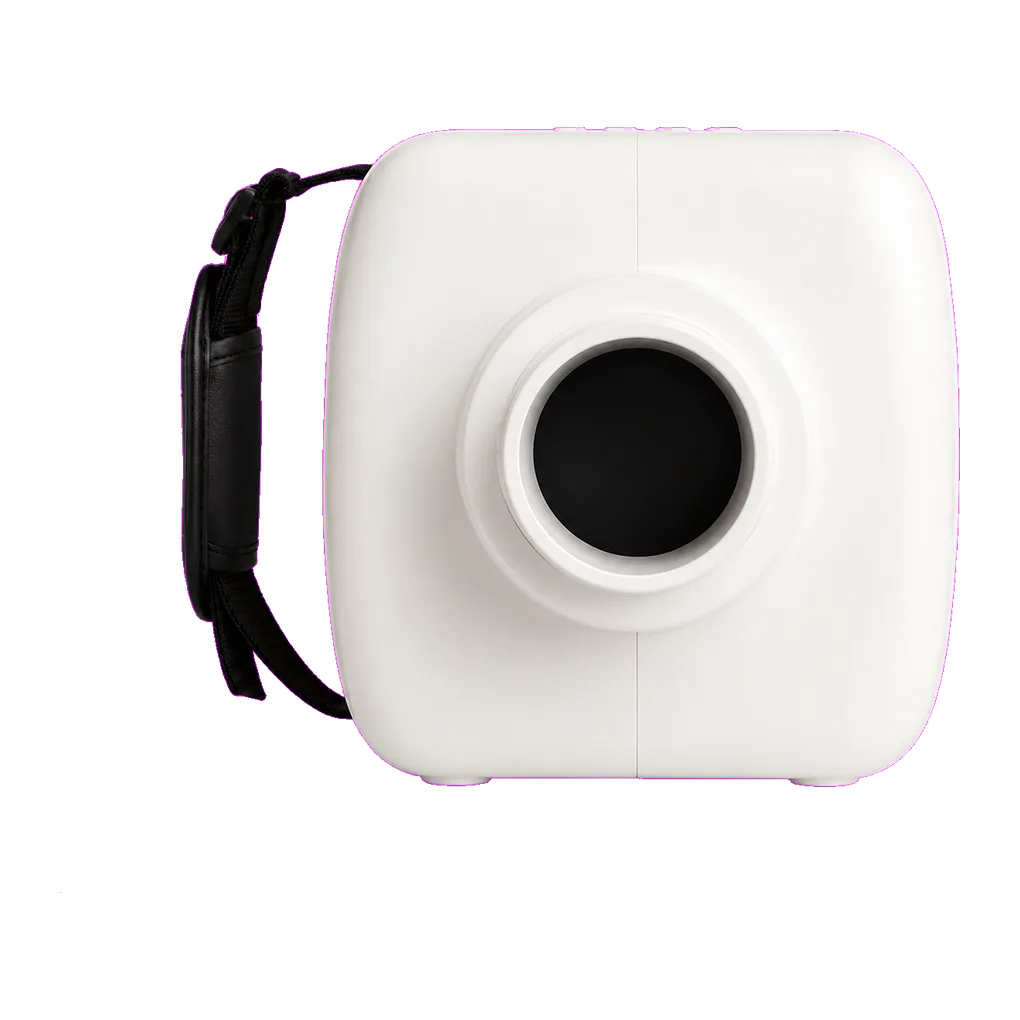
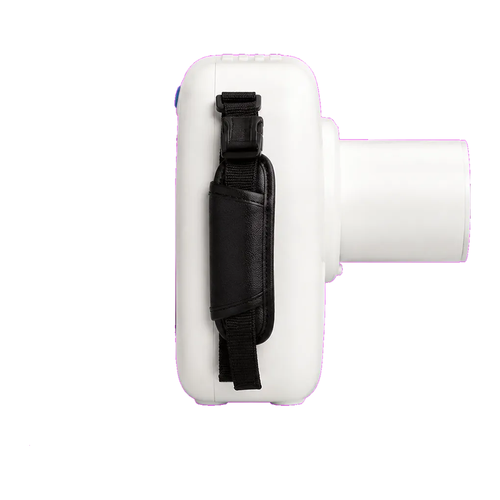
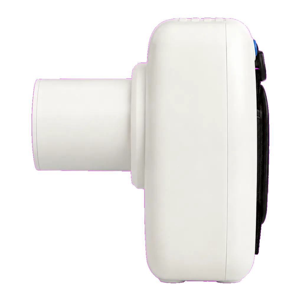
Intraoral radiography carries the diagnostic load in UK general dentistry, and the SJD-351 is configured for that daily volume. It supports bitewing caries detection, periapical assessment of pathology, endodontic length verification, alveolar bone-level review and root morphology assessment ahead of extraction.
The angle display finds its clearest use in the projections that fall outside standard paralleling technique. Paralleling with a beam-aiming holder is the recommended approach because the holder controls the geometry, and a great deal of routine work is done that way. But every practice meets patients where it cannot be applied — a shallow palate that will not accept a holder, a pronounced gag reflex, an edentulous ridge offering nothing to bite on, a young child, or a patient who can only manage a finger-held receptor. In each case, vertical angulation returns to operator judgement, and an objective figure on screen supports that judgement.
Special care dentistry, paediatric practice and domiciliary work meet those situations more often than routine general practice does, which makes this pairing of a carried unit and an angle readout a reasonable fit for those services.
Recorded angulation also supports comparison over time. Where a review radiograph is read against an earlier one — bone levels at recall, resorption through orthodontic treatment, healing after surgery — reproducing the earlier geometry keeps the comparison meaningful, and a noted figure gives the operator something definite to return to.
Multi-operator practices gain a shared reference. Where several clinicians and nurses use one unit, a recorded angulation gives the team something concrete when an image quality audit points at geometry.
Guidance from the College of General Dentistry and UKHSA Dental X-ray Protection Services remains the reference point UK practices work to, alongside HSE enforcement of IRR17.