
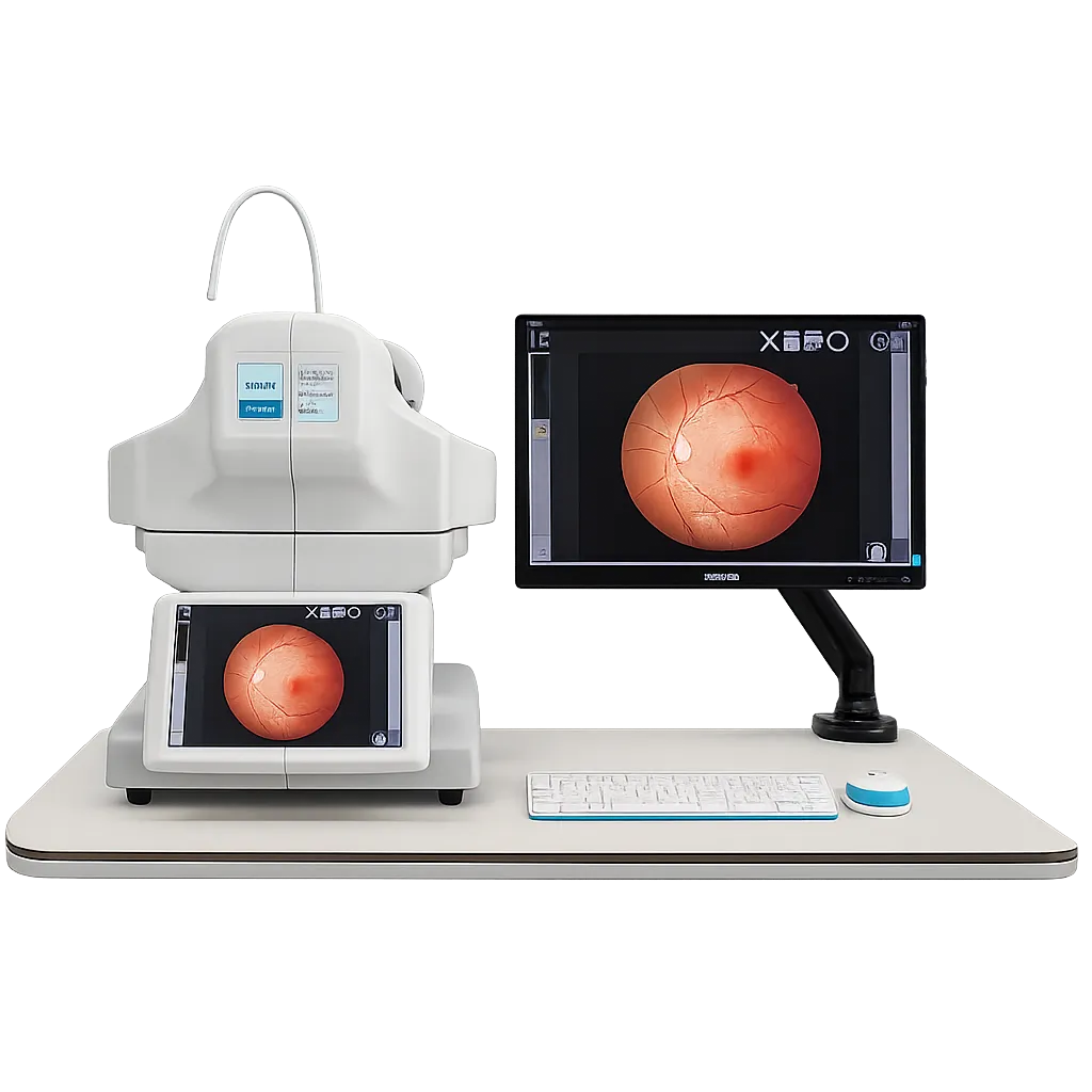
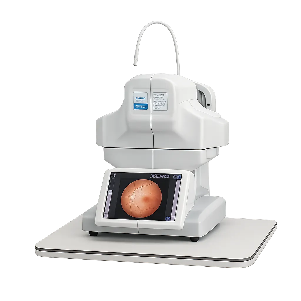
One-Key Automatic Acquisition
A single key press runs the entire sequence: chinrest adjustment, pupil search, alignment, focus, exposure and binocular capture. The operator enters patient details and the instrument does the rest.
Non-Mydriatic Design
Imaging proceeds without dilating drops, which removes the sting, the hours of blurred vision and the inability to drive afterwards that deter patients from attending screening appointments.
Minimum Pupil Size of 3.3 Millimetres
The instrument works from a pupil measuring 3.3 millimetres, so patients whose pupils constrict readily under light remain imageable without pharmacological help.
Automatic Chinrest Adjustment
The chinrest positions itself rather than requiring manual setting for each patient. Height differences between patients stop being something the operator manages between appointments.
Automatic Pupil Search and Alignment
The system locates the pupil and aligns to it independently. Accurate alignment is the part of fundus photography that usually demands practice, and here it is handled internally.
Auto Focus and Auto Exposure
Focus and exposure are set automatically for each acquisition, so image quality does not depend on how experienced the person operating the instrument happens to be that day.
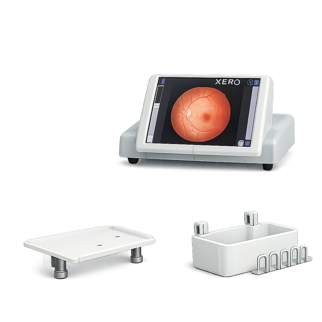
Manual Mode With Touchscreen Control
Where automatic capture cannot achieve a required view, manual operation gives touchscreen control over alignment, focus, flash and dioptre compensation.
DICOM Support
Images can travel into established hospital record and archiving systems rather than remaining on the instrument, which keeps retinal images with the rest of a patient's record.