
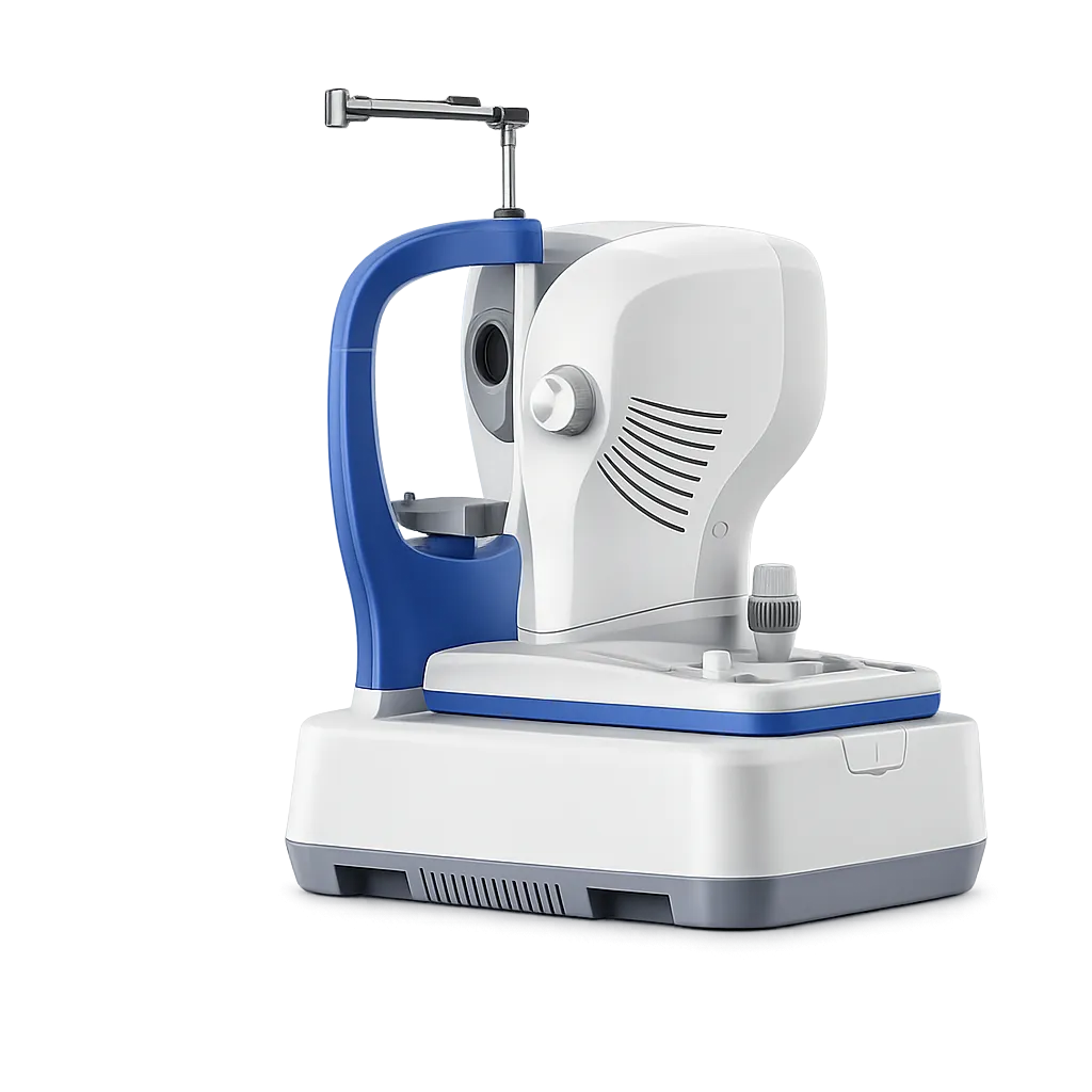
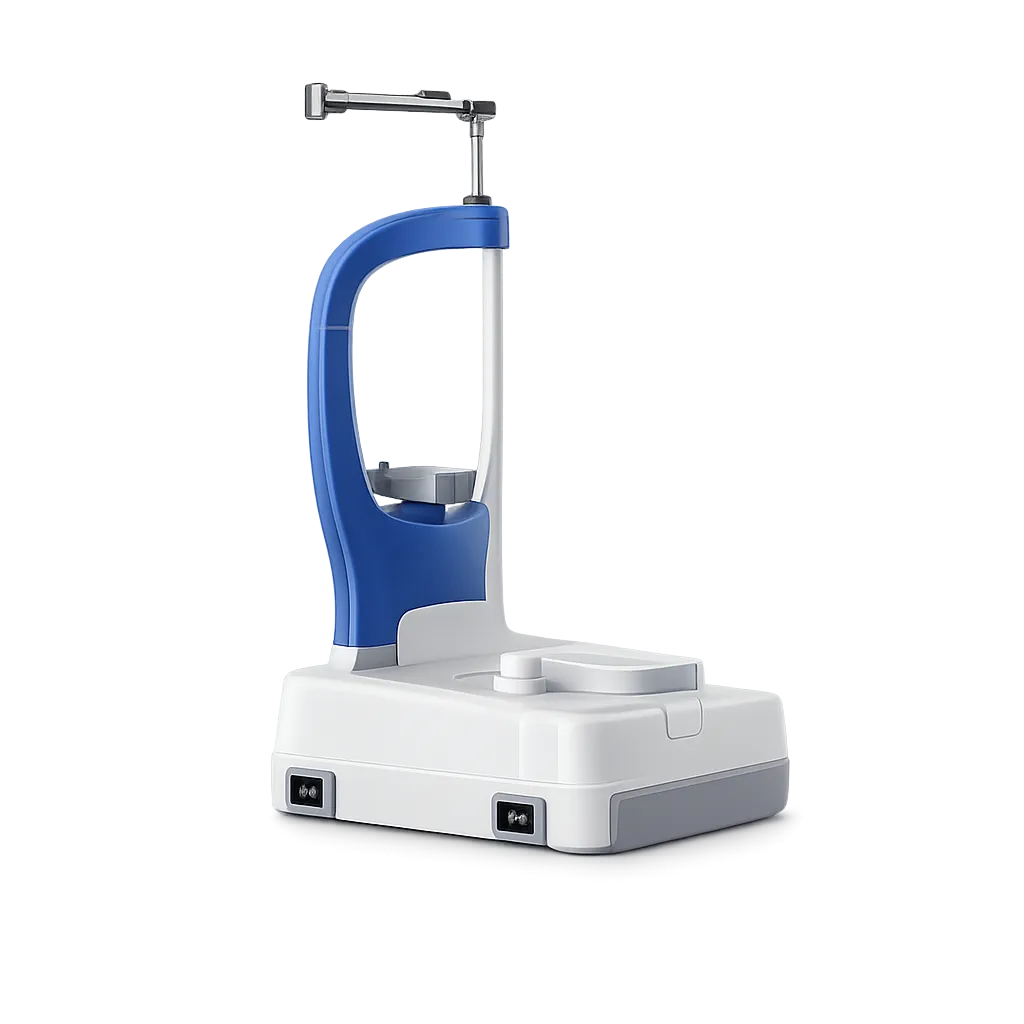
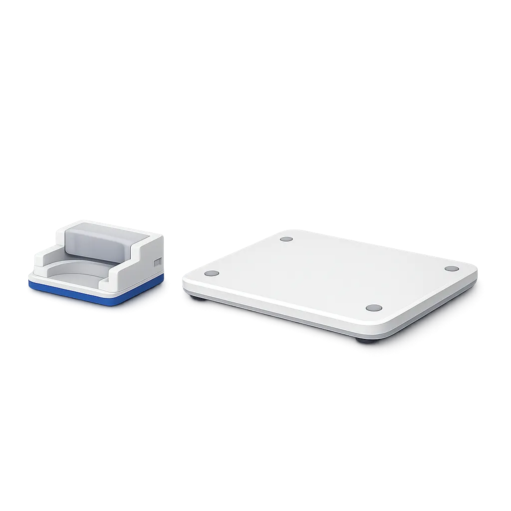
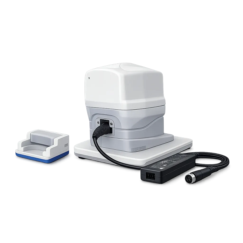
Dye Free Vascular Imaging
Angiography here is described as non invasive, studying flow within the vascular structure of the eye without dye injections. The clinical picture of perfusion is built from the scan itself.
Scan Speed of 80,000 A-Scans per Second
The stated scan speed is 80,000 A-scans per second. High speed acquisition is what makes dense angiography volumes and repeated structural patterns practical within a single sitting.
Spectral Domain Imaging from an SLD Source
Methodology is listed as spectral domain with a super luminescent diode source at 840 nm. Axial resolution is stated at 5 microns optical and A-scan depth at 3mm.
Real Time Retinal Tracking
Tracking based on laser ophthalmoscopy follows the retina during acquisition, which the manufacturer relates to reducing artefacts caused by eye movement during the examination.
Averaging Across Many Images
Ultra fine retinal imaging is described using an averaging technique of up to 50 images, so the displayed result is built from a large stack rather than one pass.
Two Angiography Configurations
The advance configuration lists four scanning volumes, eight segmentation options and quantitative analysis. The essential configuration lists two scanning volumes. Both run the C-OMAG algorithm.
Wide Field Live Fundus Imaging
Line scanning laser ophthalmoscopy provides a live fundus view across a 45 degree field, with a minimum pupil diameter of 3.0mm listed for the imaging.
Full Structural Pattern Set
Macular, disc and anterior patterns are all listed, alongside two glaucoma cubes sampling evenly across 200 by 200 A-scans over the macular area and the disc area.