
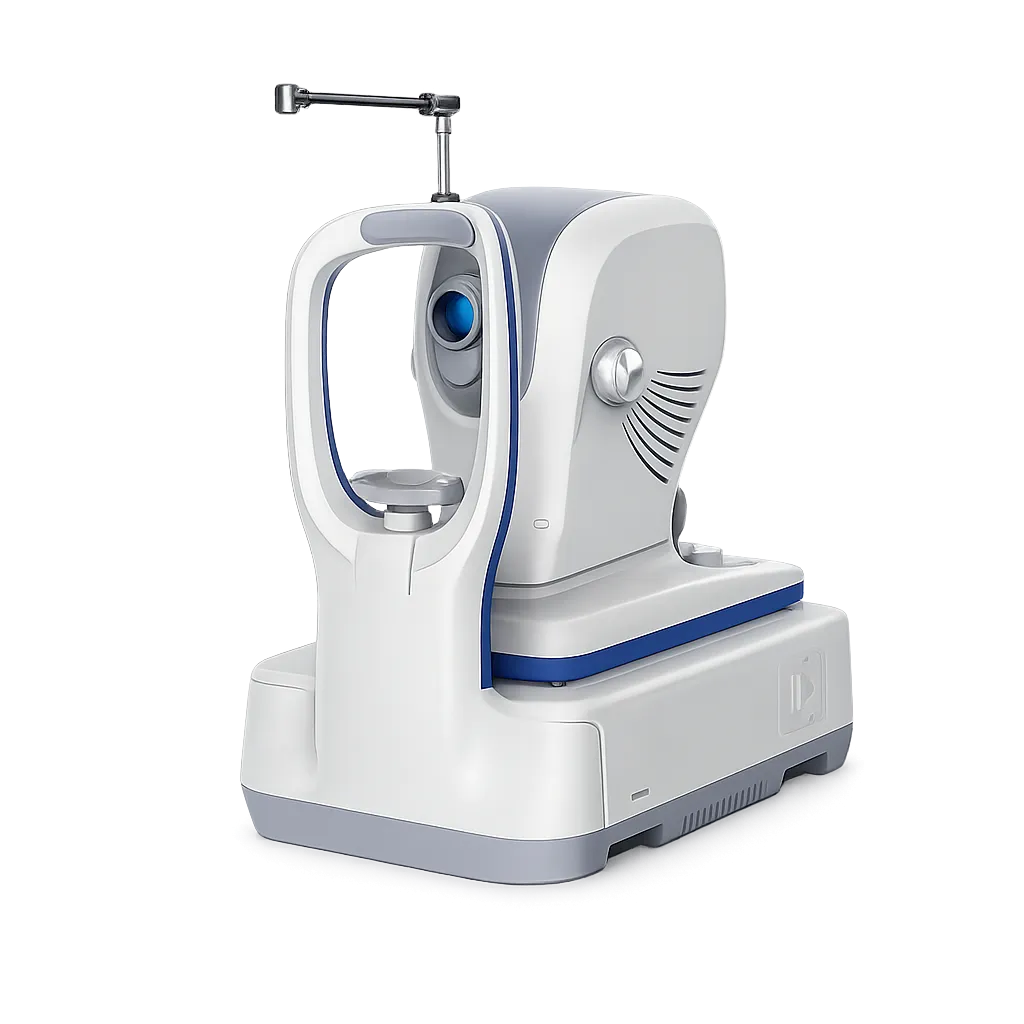
Spectral Domain Methodology
The methodology is listed as spectral domain, working from an optical source with a light centre wavelength of 840nm. A-scan depth is given as 3mm, which sets how far into the tissue each layered image extends.
Five Micron Optical Axial Resolution
Axial resolution is stated at 5 microns optical and 3.6 microns digital, with transverse resolution at 15 microns optical and 3 microns digital. Those figures govern how finely separate tissue layers can be told apart.
Two Glaucoma Cube Patterns
The data describes a glaucoma cube over the macular area and a second over the disc area. Sampling is evenly distributed across 200 by 200 A-scans, which the manufacturer relates to early detection.
Dense Retinal Thickness Cube
Retinal thickness is assessed point by point across a dense 500 by 100 cube. That density supports the thickness maps and progression comparisons clinicians rely on when following a patient over years.
Anterior Segment Line Scanning
The data lists an HD line scan at 6 or 16 mm and a 6-line radial scan for the anterior segment. Corneal imaging at this definition locates Bowman's layer, between stroma and epithelium.
Laser Fundus Imaging
Fundus imaging uses line scanning laser ophthalmoscopy, with a minimum pupil diameter of 3.0mm and a field of view of 40 by 30 degrees, so a surface view accompanies the layered study.
Wide Dioptre Compensation
The dioptre range runs from minus 20 to plus 20, covering a broad span of refractive error without separate correction at the instrument.
DICOM Conformance and Remote Viewing
DICOM conformance is listed, and remote viewer software is noted as available, so studies can be handled within the imaging arrangements a clinic already runs.