
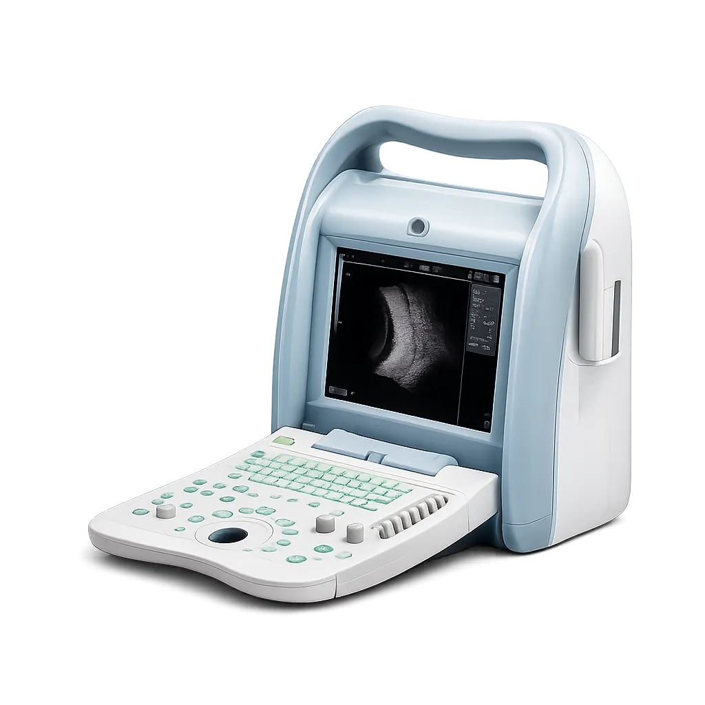
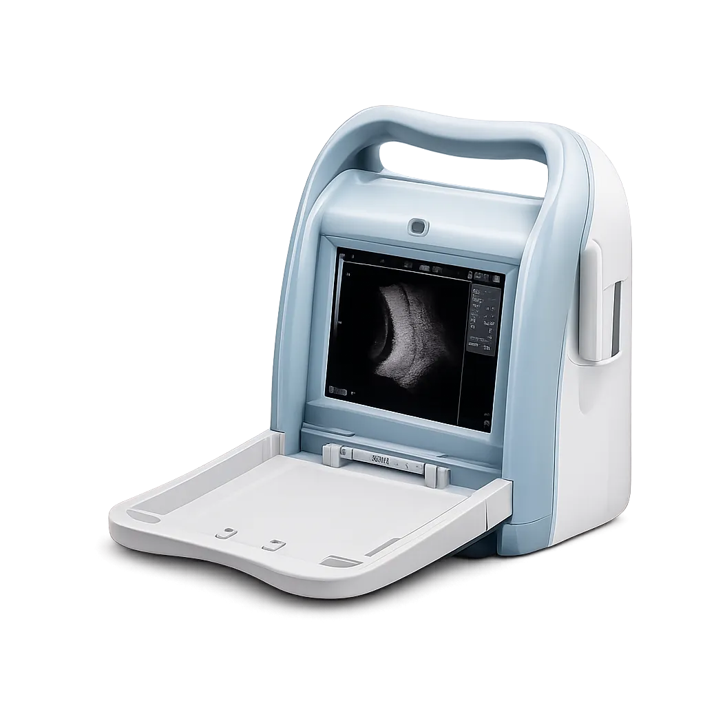
Hospital ophthalmology departments are the setting this model is built for, and DICOM 3.0 is why. A department running a PACS needs imaging studies to be entered into the record automatically rather than printed or exported by hand, and a scanner without DICOM sits permanently outside that workflow. Password-protected presets and named system configuration further suit a device shared across a department rather than one owned by a single clinician. Medigear.uk verifies certifications and confirms network configuration requirements before any procurement decision is made.
Posterior segment assessment benefits from the width of the image controls. Gain across 0 to 120 d, B with separately adjustable dynamic range and eight-level time gain compensation, gives more latitude than compact units typically offer, which matters when distinguishing vitreous opacity from retinal detachment,t where reflectivity differs subtly. Five display modes and a measurable 256-frame cine loop support review of dynamic findings rather than still frames alone.
Cataract and IOL planning uses A-scan with both single-point and five-point measurements to measure anterior chamber depth, lens, vitreous, central corneal thickness ,ness and axial length by immersion. Immersion avoids the corneal compression contact applanation introduces, and five-point measurement supports the repeat-reading discipline biometry requires. All interpretation remains with the practitioner.
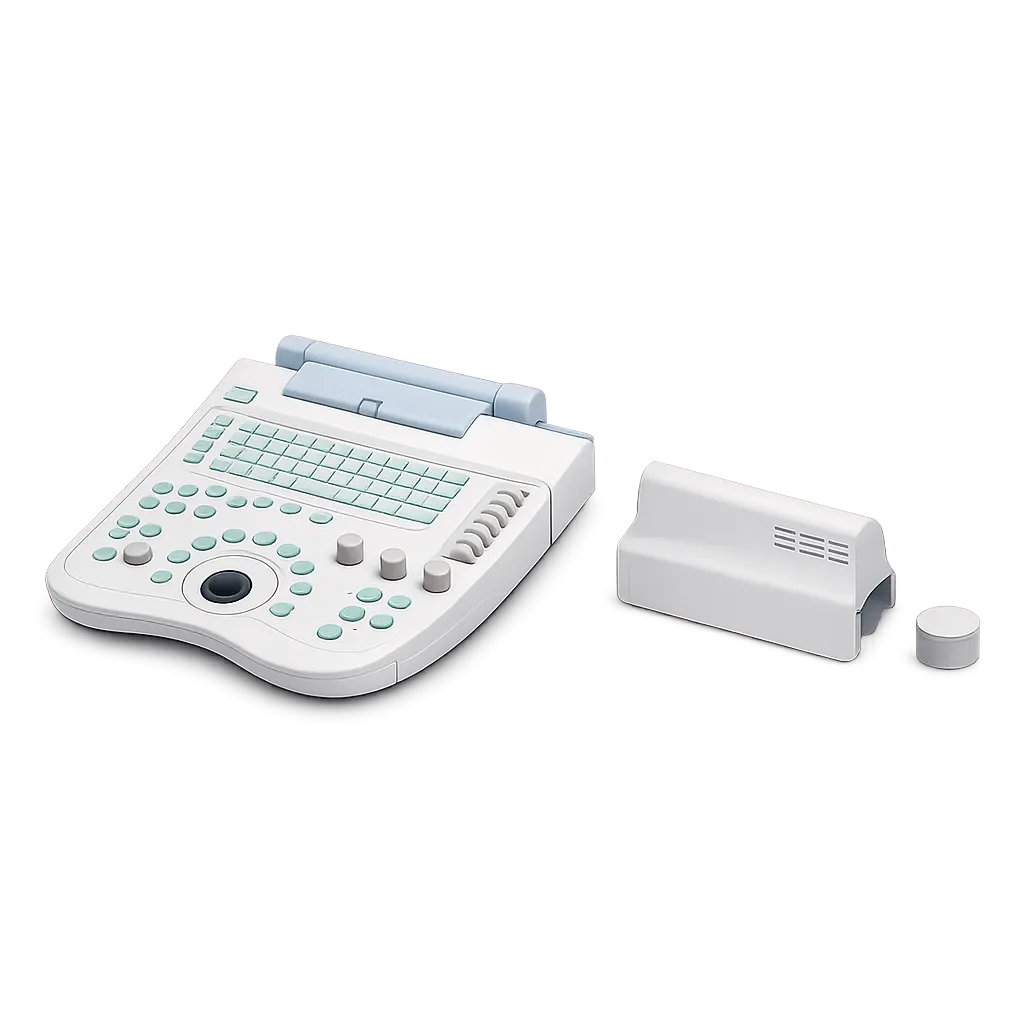
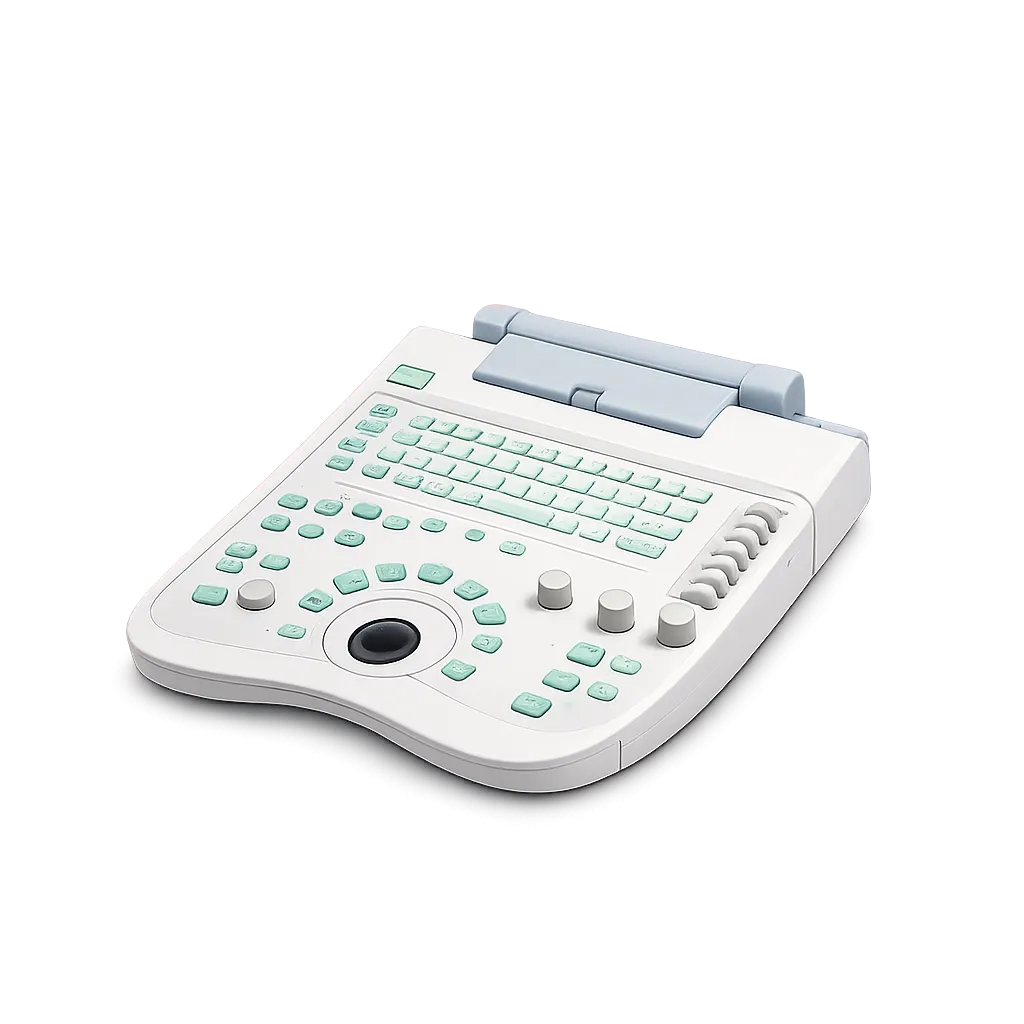
Departments examining in reduced light gain from the backlit silicone keyboard with trackball and encoder. B-scan is routinely performed with the room dimmed, and an unlit console forces either working by feel or raising the lights. Medigear.uk supplies the operating documentation with the unt, and confirms probe supply and replacement availabilitin theat quotati,on so a department knows the ongoing position rather than only the purchase price.