
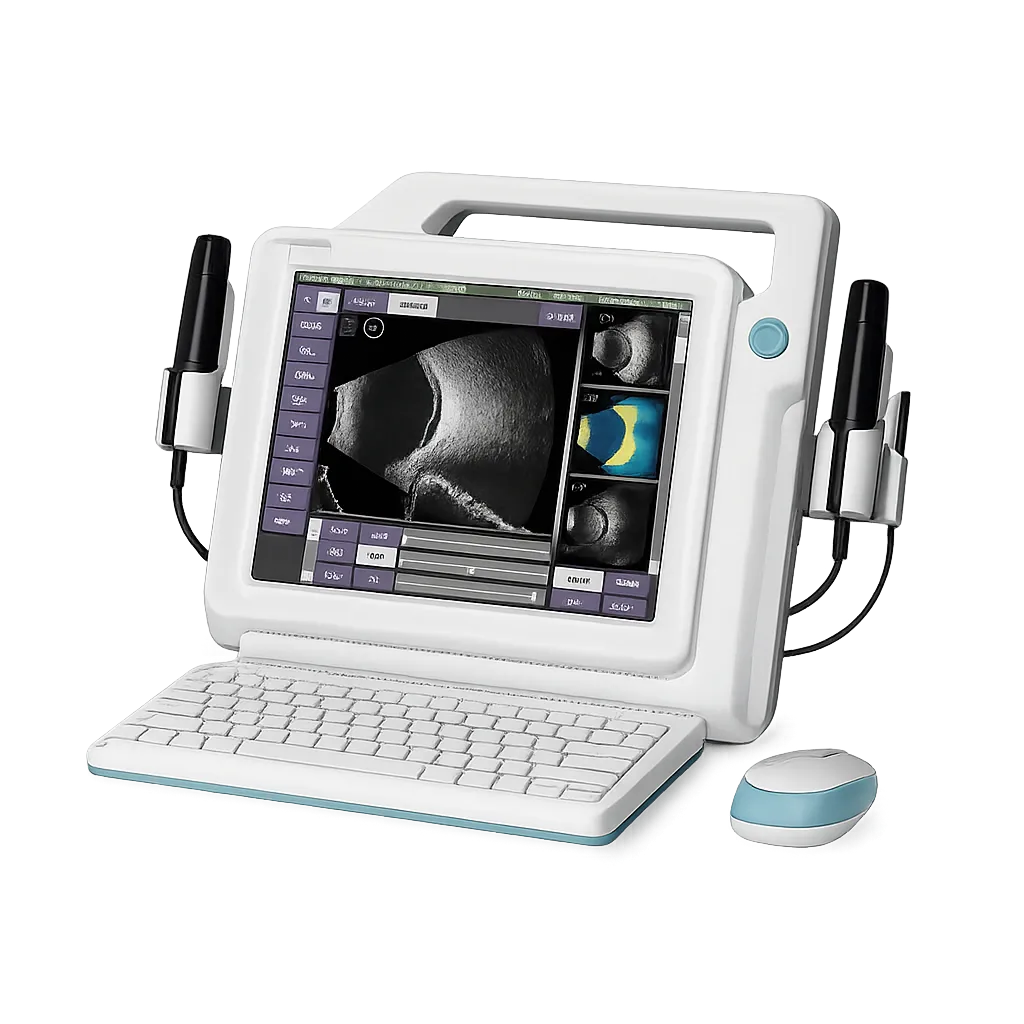
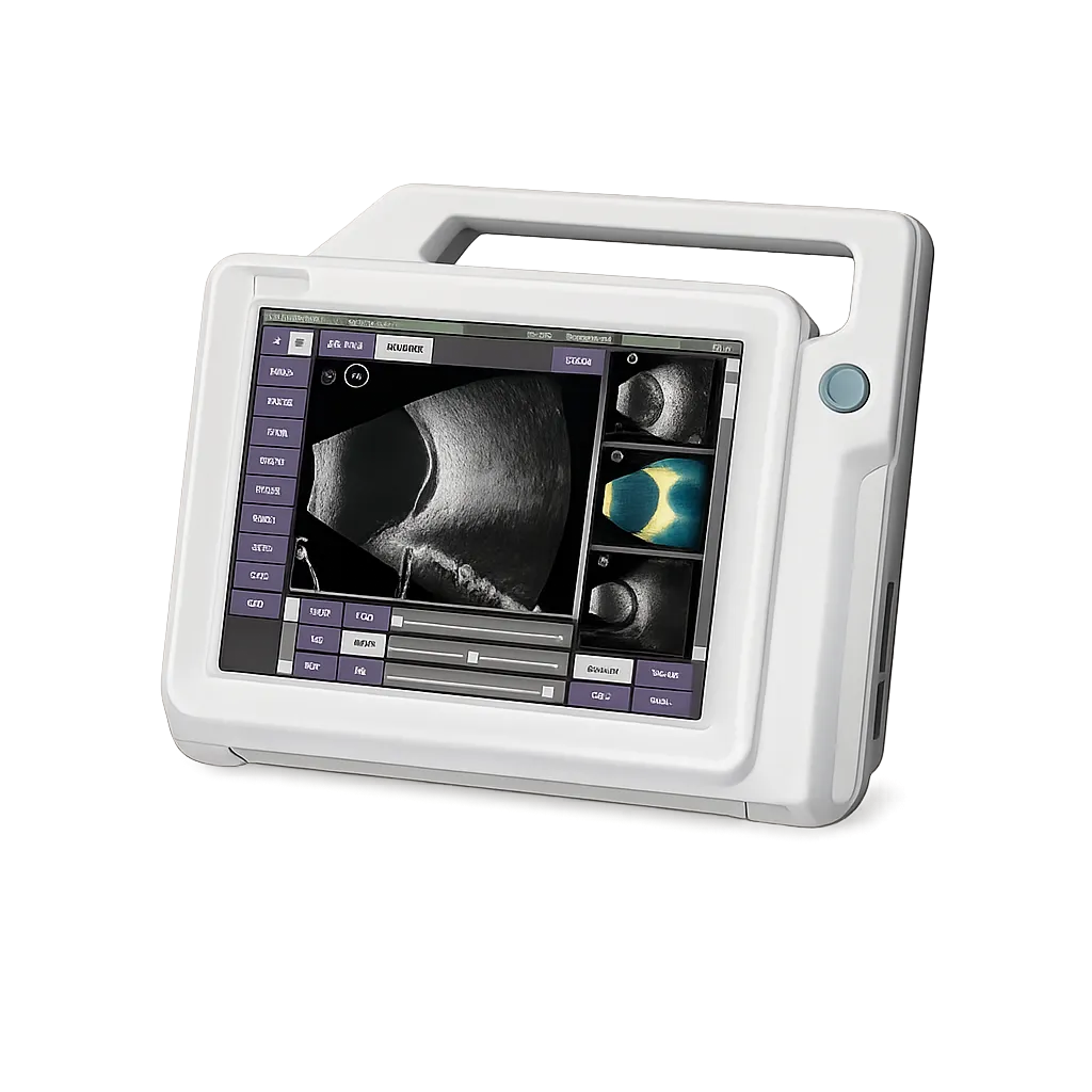
Ophthalmology clinics assessing the posterior segment gain most from having both frequencies available. The 10 MHz probe reaches 60 mm and renders low-reflectivity vitreous well, which is what distinguishes vitreous haemorrhage from retinal detachment. The 20 MHz probe resolves to 0.08 mm axially at shallower depth, showing fine membranes and shallow detachments that a lower frequency renders less distinctly. The manufacturer's clinical gallery presents the same pathologies at both frequencies, which is the clearest illustration of why one probe is a compromise. All image interpretation remains with the practitioner.
Anterior segment work benefits directly from the 20 MHz probe over a 19 to 40 mm range, where penetration is not the constraint and detail is. Practices that would otherwise refer such assessments out or accept lower-resolution imaging can perform the work in-house instead. That keeps the patient in one appointment and the finding in one record.
Cataract planning uses biometric A-scan with IOL formula calculation, and the optional immersion shell is worth specifying where axial length accuracy matters, since immersion avoids the corneal compression that contact applanation introduces. Medigear.uk prices the shell alongside the base unit and confirms the IOL formula set at quotation, since formula availability determines which eyes a surgical service can plan for.
Services where several people examine benefit from reporting built on a configurable dictionary rather than free text. Consistent terminology across operators makes findings comparable between appointments and clearer on referral. Multi-buffer capture also allows two frequencies or two eyes to be compared during the appointment rather than afterwards, and Medigear.uk confirms buffer count with the manufacturer before order.