
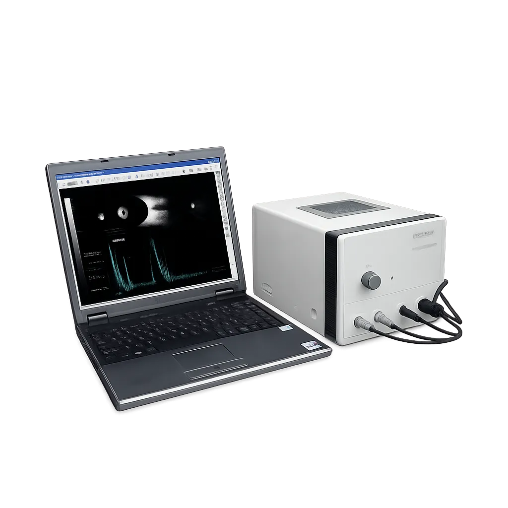
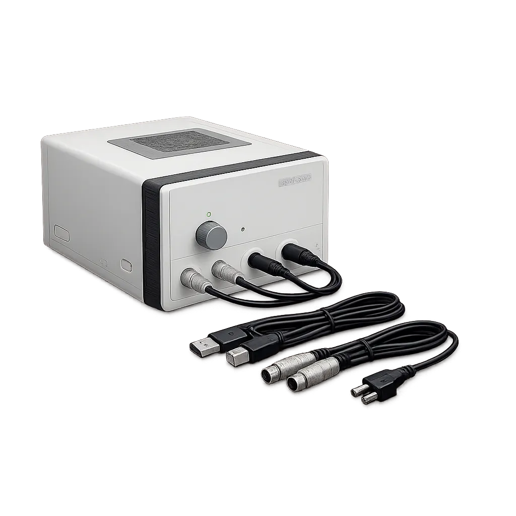
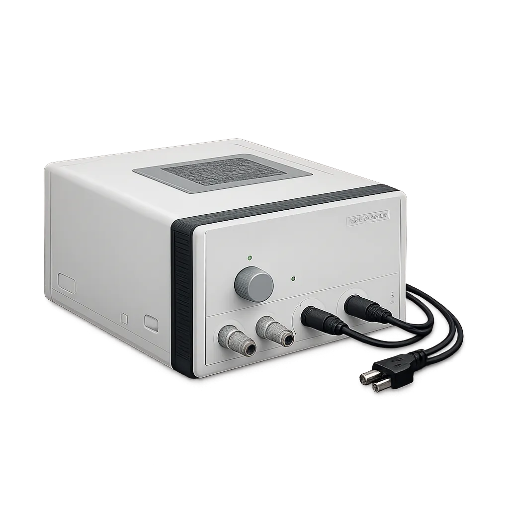
Ophthalmology clinics use B-scan where the fundus cannot be viewed directly, and this model addresses a specific frustration in that work. A B-scan examination is a sweep, and the frame that most clearly shows a finding is often not the one the operator froze on. Dynamic playback allows the sequence to be reviewed and the best frame selected afterwards, so the examination is judged on its clearest image rather than its last. That also reduces the need for repeat sweeps, which matters for anxious or uncomfortable patients. All image interpretation remains with the practitioner.
Services building an evidence record benefit from images being delivered to the workstation as they are acquired rather than through a separate export. An image captured but never filed is a familiar failure in busy clinics, and removing the manual step removes the opportunity for it. Where a service must provide evidence for referral or audit, the completeness of the record matters as much as image quality. Medigear.uk confirms how the workstation stores, exports, nd prints records before any procurement decision is made is made
Posterior segment assessment depends oon grey-scaledepth, since distinguishing vitreous opacity from retinal ddetachment relies on resolvingsmall differences in reflectivity. The manufacturer states 256 levels against 64 in a single-chip architecture, alongside probe gain from 30 dB to 105 dB, vitreous and retinal enhancement, and multi-colour false colour.
Cataract planning uses A-scan measurements of anterior chamber depth, lens thickness, and axial length, along with implant power calculation. Because the CAS-2000 designation covers several variants with differing capability, Medigear.uk confirms the A-scan specification and formula set for the exact variant quoted rather than relying on a family listing.