
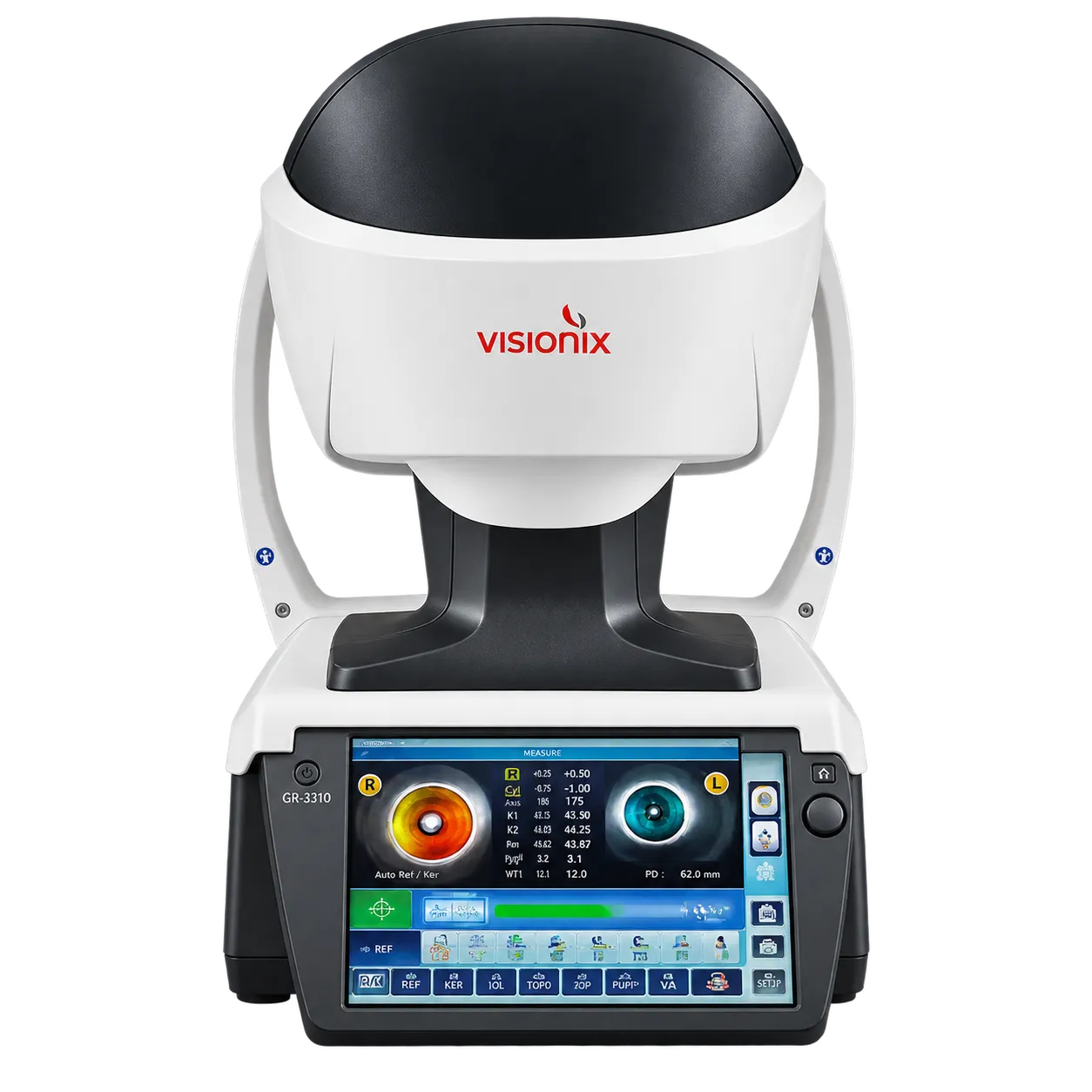
Glaucoma services get structural and pressure data from the same examination. The VX 130+ reports the iridocorneal angle and anterior chamber volume, along with IOP and pachymetry-weighted IOP, so the pressure figure reflects corneal thickness. Angle and chamber volume describe the drainage anatomy, while weighted IOP reflects pressure. Repeat examinations then build a record for monitoring over time.
Keratoconus patients and contact lens wearers form a second group. Continuous keratoconus monitoring supports fitting rigid and ortho-k lenses, allowing clinicians to see corneal changes between fittings. Contact lens fitting is a one-step process. The VX 130+ also screens for corneal pathologies, with corneal topography in the same instrument as the Scheimpflug camera.
Cataract and refractive surgery candidates gain planning data. Posterior corneal keratometry feeds into IOL calculations, and the kappa angle is considered to avoid post-surgery decentration. Lens opacities can be visualised during the same visit. Objective refraction and quality-of-vision analysis cover day and night conditions, with sphere from −20 to +20 D and cylinder up to +8 D. The refraction data are completed by an axis range of 0 to 180° and a measuring range of 2 to 7 mm in three zones.
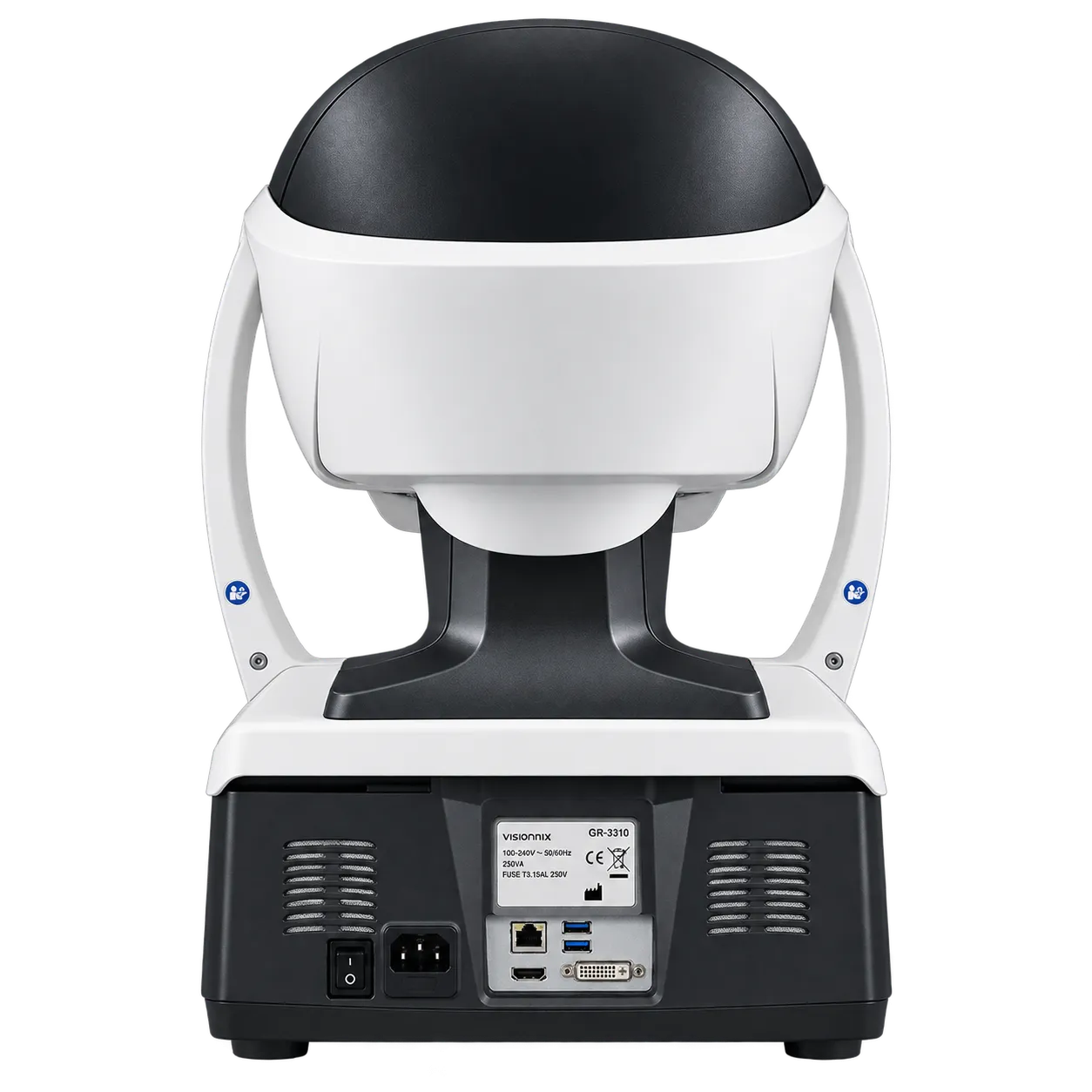
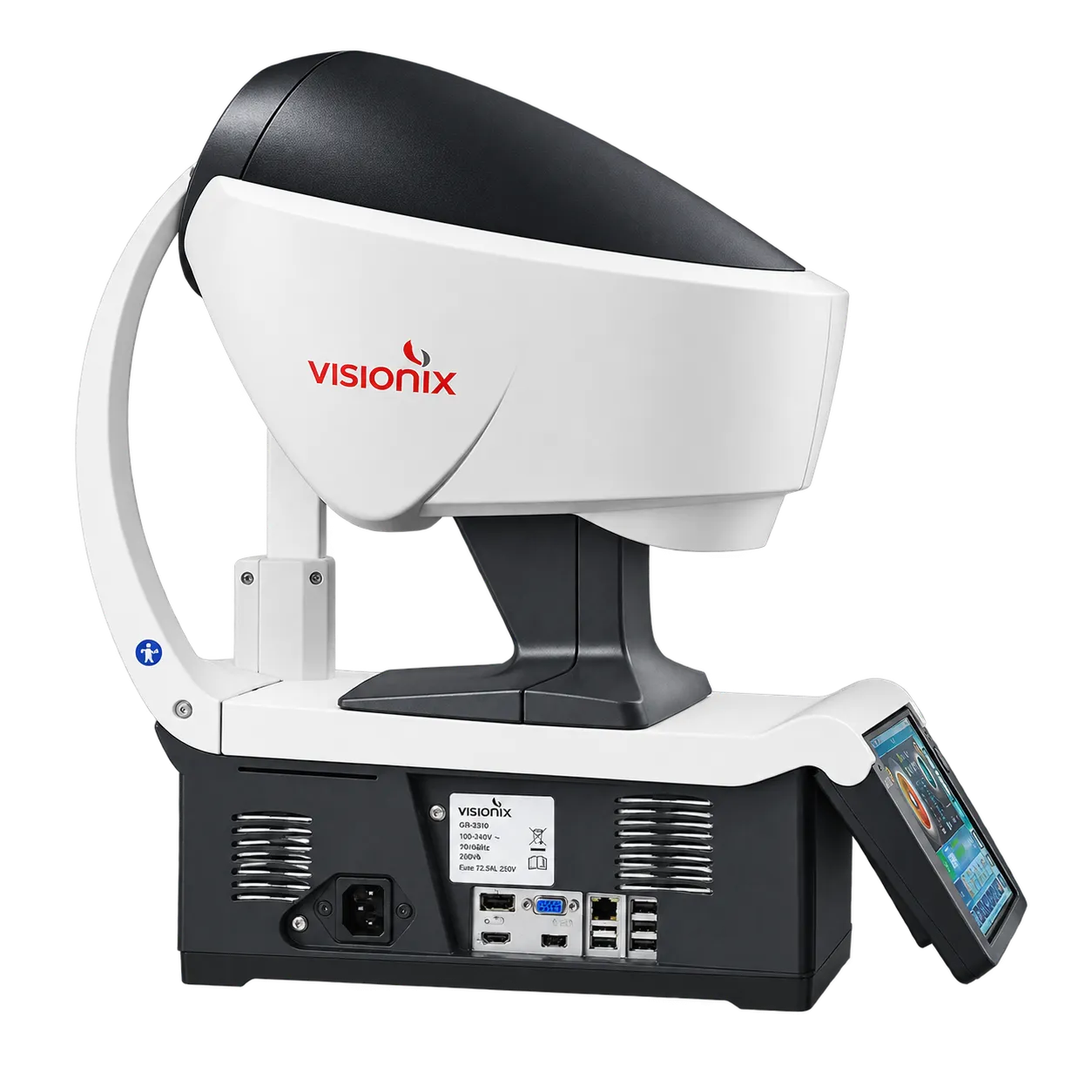
Because the examination is fully automated and delegable, staff other than the examining clinician can run it while the clinician reviews the results. Installation requires a table that supports 25 kg, a 100–240 V supply, and space for a 312 × 530 mm footprint. Retinal imaging and subjective refraction fall outside its scope, so those steps need other instruments.