

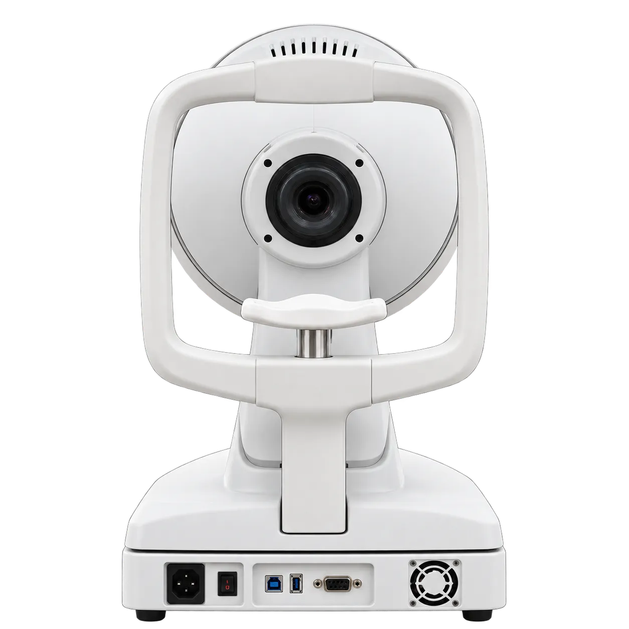
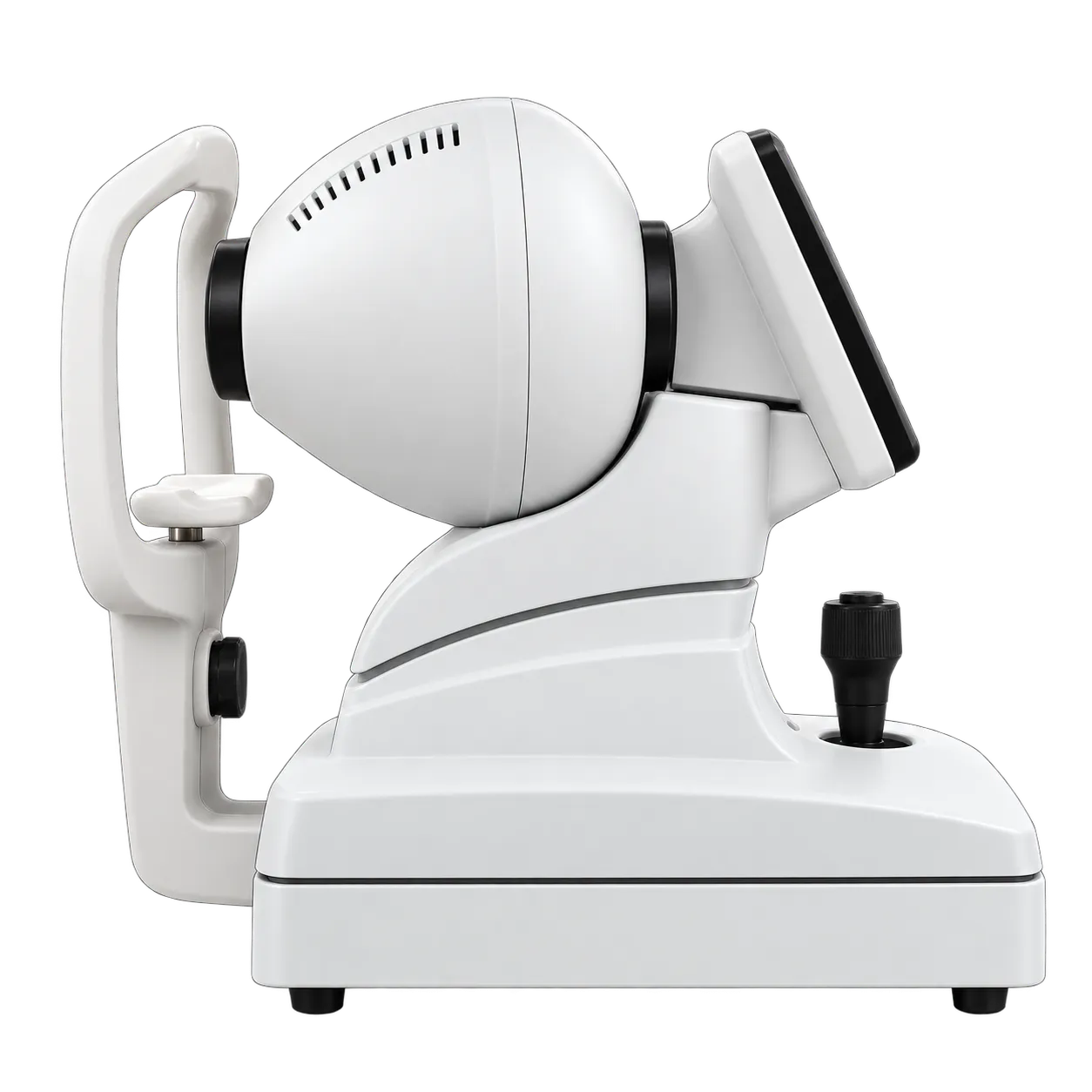

The CA-800 fits ophthalmology clinics, optometry practices, contact lens fitting rooms and dry eye services. An operator follows the visual guidance for alignment and focus, and the auto shot takes the image once the eye is in focus. Right and left eye results appear on one screen, which keeps a visit's data together for the clinician. A 10.1-inch capacitive touch screen runs the exam, and the integrated PC holds the patient database, so capture, review, and reporting happen at one station.
For corneal assessment, the topography, 3D, height, comparison and differential maps show curvature and elevation across the 9.8 mm coverage. The keratoconus probability index shows a green, yellow or red result, and the Cone Location and Magnitude Index supports follow-up at later visits. Zernike analysis adds corneal wavefront detail with 36 polynomials up to the seventh order.
Contact lens work uses the simulation software and the lens database, and fluorescein imaging and video with eight blue LEDs show the fit in real time. Pupillometry records pupil position and diameter in dynamic, photopic, mesopic, and scotopic conditions, with a response-latency graph.
Dry eye assessment draws on infrared meibomian gland analysis, blink detection and tear break-up time. White-to-white measurement and ring editing support routine corneal work, and the comparison and differential maps let clinicians compare one exam with an earlier one at a follow-up visit. Reports leave the instrument as PDF or print, and the i-Map option lets support staff acquire data while the clinician reviews it in another room.