
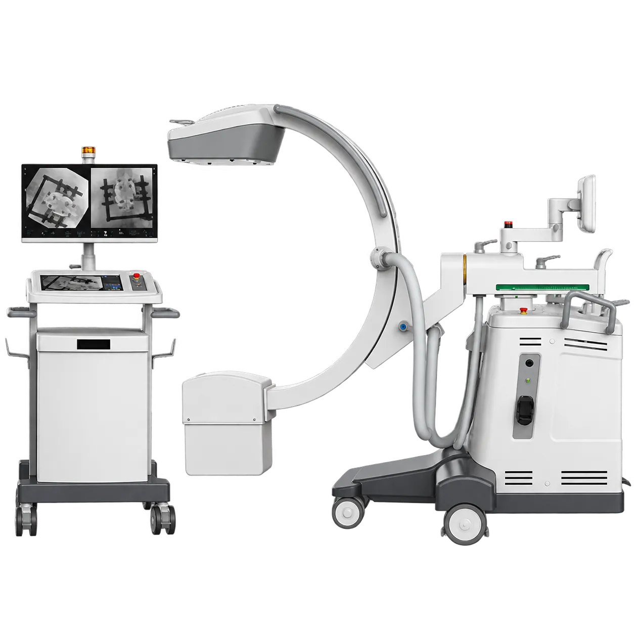
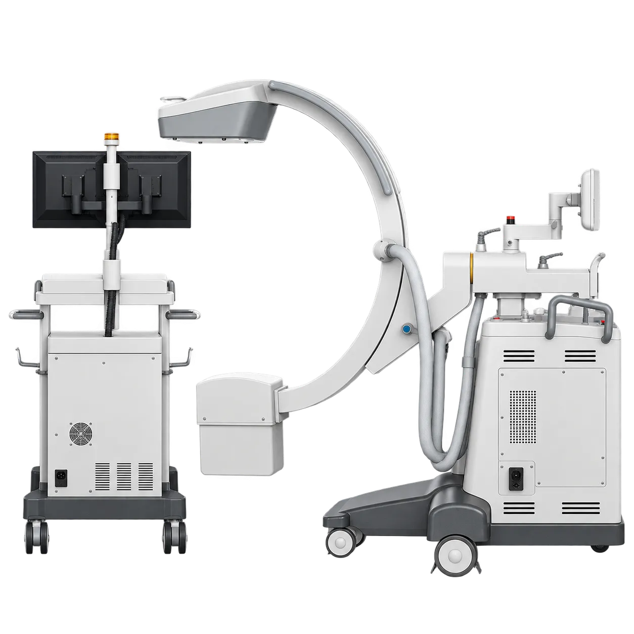
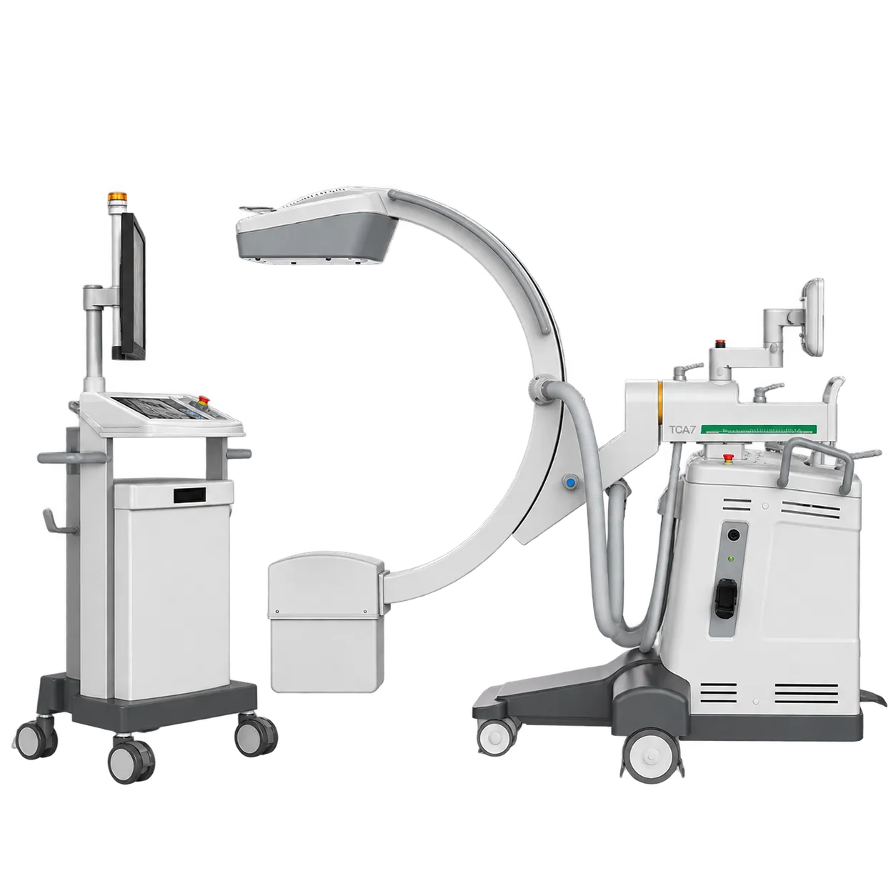
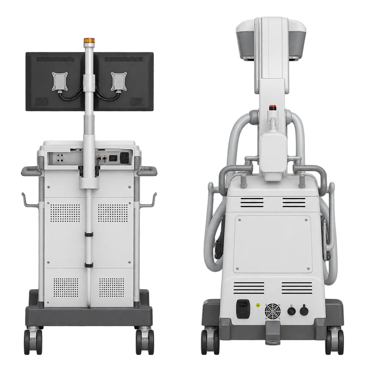
Spine fixations show what the water cooling is for. These are long cases with fluoroscopy at intervals from start to finish, and the cooled monobloc keeps the generator working throughout. Orbital rotation of 169°, with overscan, gives the surgeon a wide choice of projections. Orthopaedic theatres use the same stand for fracture and joint surgery, where metal detection keeps prostheses out of dose control so the image stays balanced around the implant.
Cardiac work draws on the same endurance. Pacemaker implantation and electrophysiology procedures run live and reference images side by side on a split-image monitor up to 42.5 inches. Cardiac and vascular surgery calls on DSA (digital subtraction angiography) in the imaging software, and Lens Boost raises contrast in a chosen region of the image.
Gastroenterology teams use the TCA 7 for ERCP (endoscopic retrograde cholangiopancreatography), pain specialists for pain management and urologists for urological procedures, each starting from APR programmes set for the anatomy and application. For paediatric applications, the anti-scatter grid comes off, both collimators offer motorised paediatric filters, and the IGZO detector option improves image quality at low dose.
Surgeons, cardiologists, gastroenterologists, and pain specialists lead these procedures; radiographers and theatre nurses run the system; and medical physicists use the dosimetric information it displays and stores for each exam. During acquisition, the support arm holds the image monitor in view while the operator stays outside the sterile field. After the examination, the 130 kg display station can be used on its own. What ties these settings together is duration: the TCA 7 is built to keep imaging through procedures with long exposure times.