
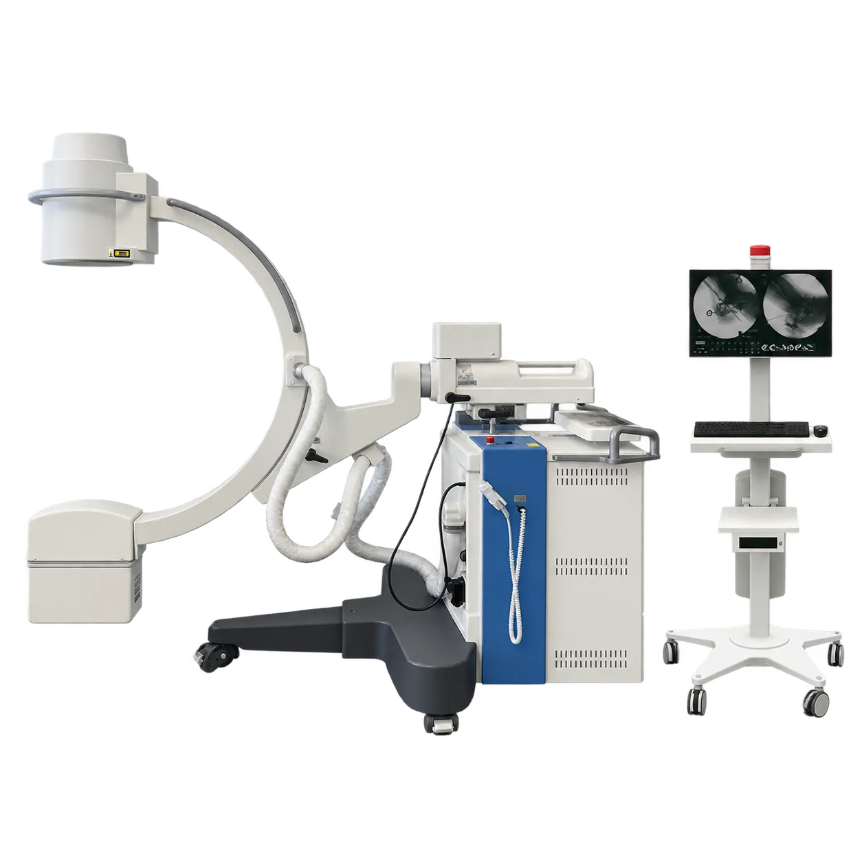
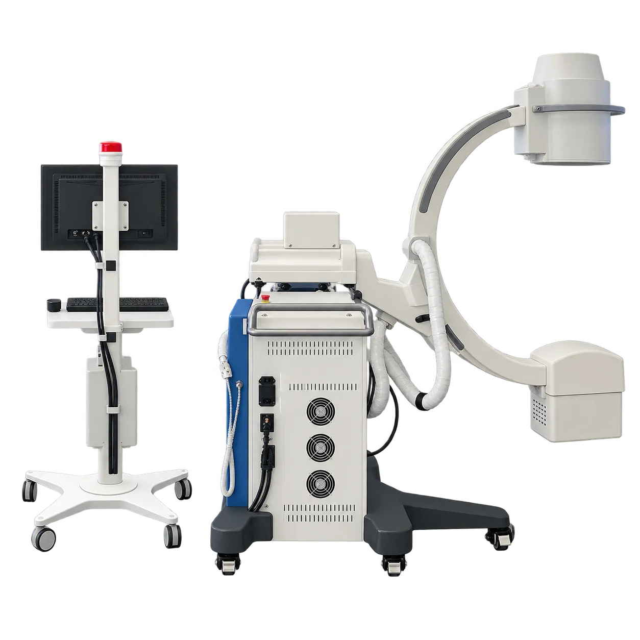
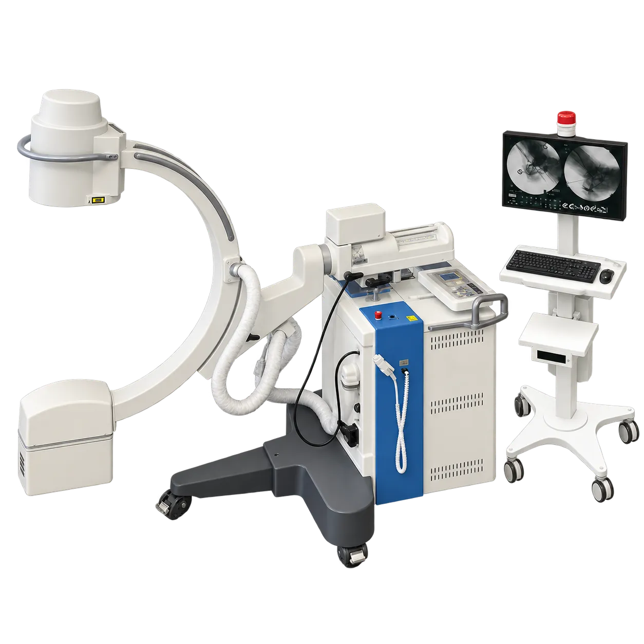
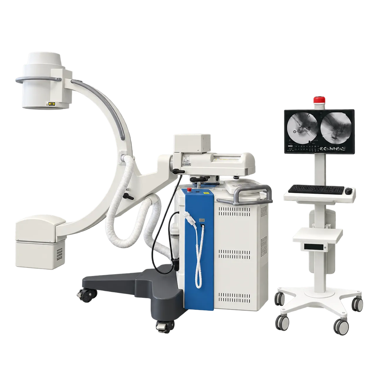
Orthopaedic and trauma theatres are a natural home for the TCA 6. During fracture reduction and fixation, the surgeon follows reduction and implant position on the trolley monitors across the table, while the radiographer moves the C-arm between anteroposterior and lateral views. Horizontal exposures through the wide orbital rotation give the lateral view without moving the patient.
Vascular surgery draws on the DSA package. Road mapping with maximum-opacity acquisition, subtraction, auto mask, pixel shifting, and manual stenosis measurement supports peripheral vascular procedures, with two 19-inch monitors or a single 24-inch monitor showing the images where the team can see them.
General surgical lists in hospitals and day-surgery units use the same set-up for intraoperative imaging wherever a 9-inch field covers the anatomy. The large distance between the stand and the beam axis leaves space around the C-arm, so anaesthetists and scrub staff can maintain access to the patient.
Dose management fits into the routine. The laser targeting device and virtual collimator set position and field without exposure, and departments with the optional dose meter keep DAP, RAK, RAKR, and effective time on record for each case, which medical physics teams can review.
Its users are surgeons, radiographers, theatre nurses and the medical physicists who oversee dose. In each setting, the separate trolley means the display goes where the surgeon looks, and the stand goes where the patient lies, and the optional wireless footswitch leaves the floor between them free of a pedal cable. Quick-clean surfaces and fast sterile cover changes keep turnaround short between cases.