
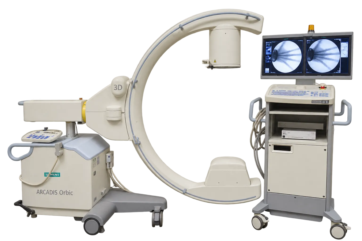
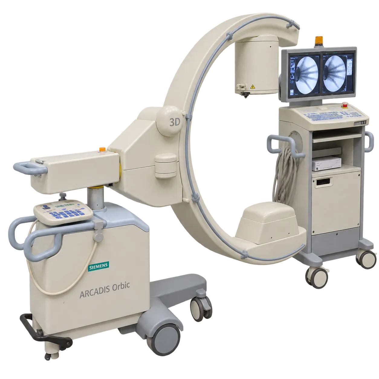
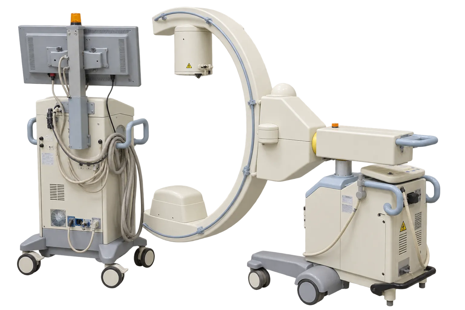
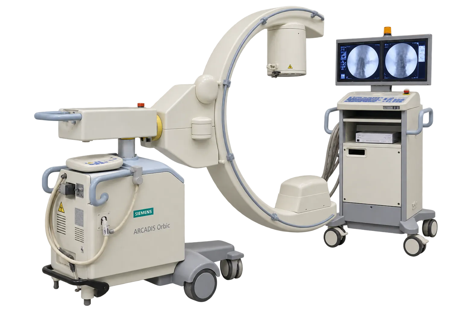
Spinal surgery is the application the three-dimensional capability was built for. Conventional fluoroscopy provides anteroposterior and lateral views, and a medial pedicle screw breach is best appreciated in the axial plane, which two-dimensional imaging does not produce. A volumetric dataset acquired in theatre provides that plane while the patient is still anaesthetised, so a finding can be acted upon in the same session rather than after a postoperative scan. Siemens states the capability reduces misplacement and revision rates. All clinical judgement, including whether an exposure is justified and what any finding warrants, remains with the operating surgeon, and Medigear.uk supplies configuration and condition detail rather than clinical guidance.
Trauma surgery uses the same capability across joints of the upper and lower extremities, pelvis and hip, where articular reduction is assessed on a volume rather than inferred from two projections. Because acquisition can be repeated during the procedure, a reduction can be checked, adjusted and checked again rather than committed to on the strength of two views. Medigear.uk confirms the 3D function is present and calibrated on any unit quoted.
Maxillofacial work is listed among the applications, benefiting from the same volumetric assessment in a region where anatomy is complex and individual projections are difficult to interpret in isolation. Medigear.uk confirms which anatomical programmes are fitted on a given machine.
Two-dimensional work benefits from the isocentric geometry alone. Because anatomy stays centred through 190 degrees of orbital rotation, changing projection needs no repositioning, which Siemens describes as saving time and dose since repositioning under screening is itself exposure.