
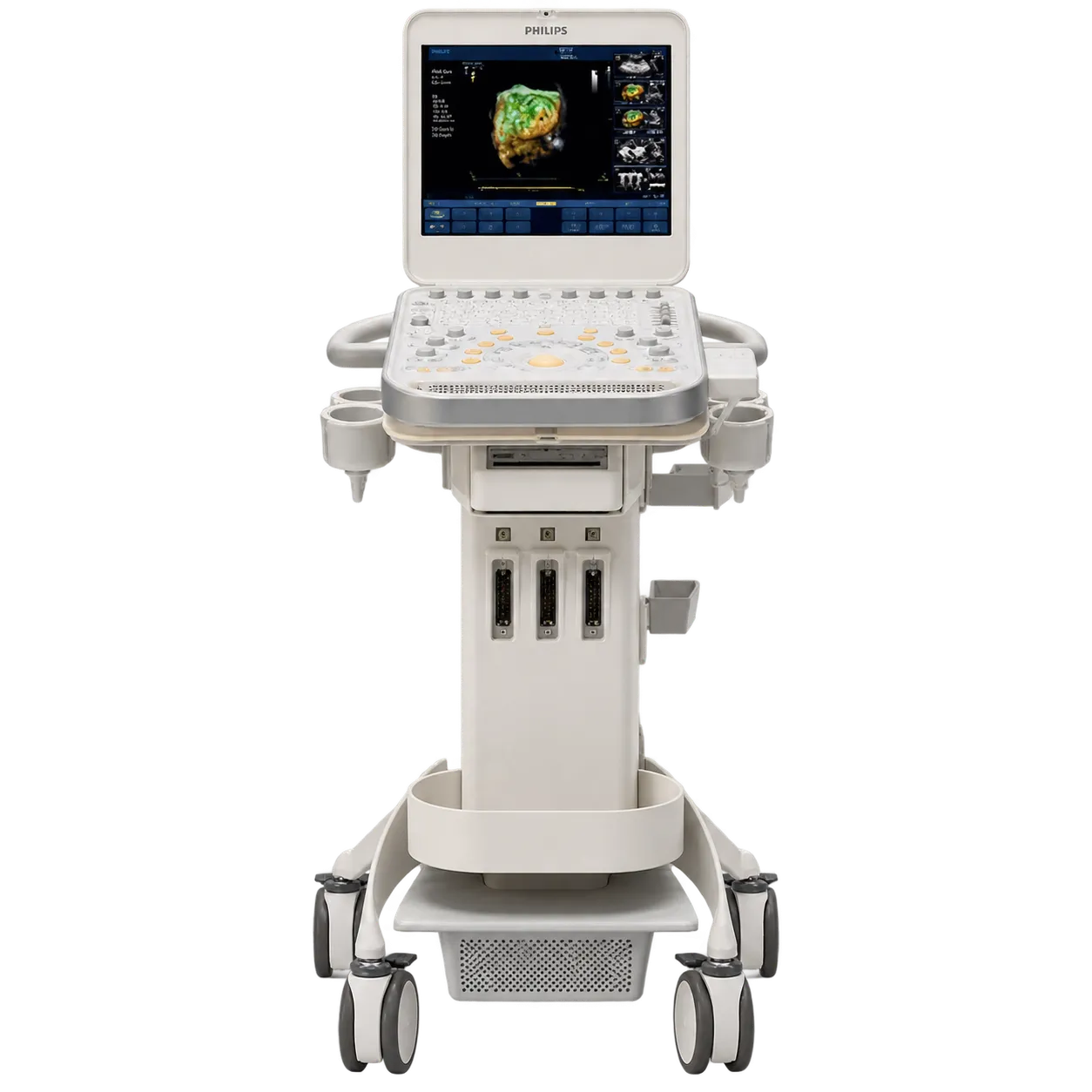
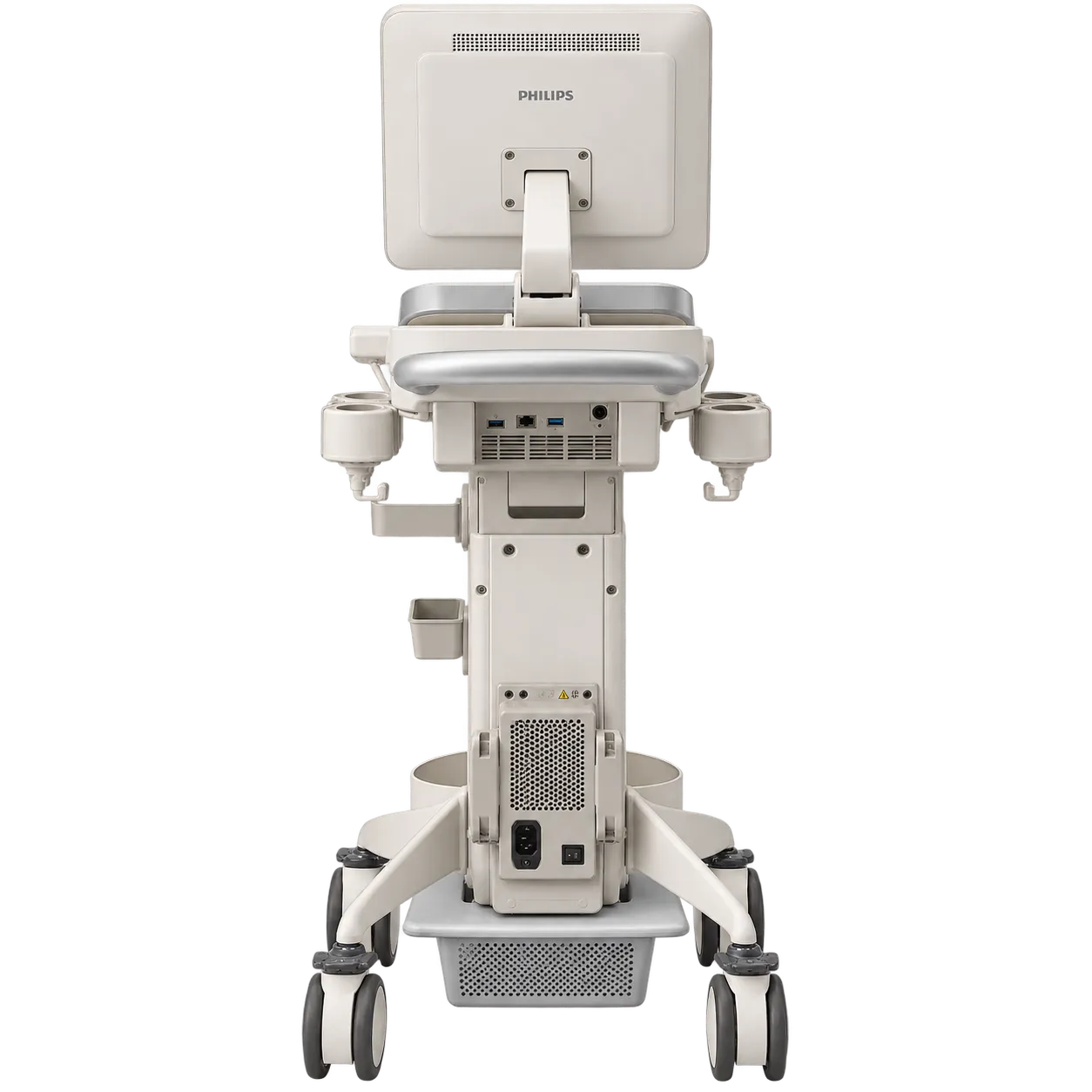
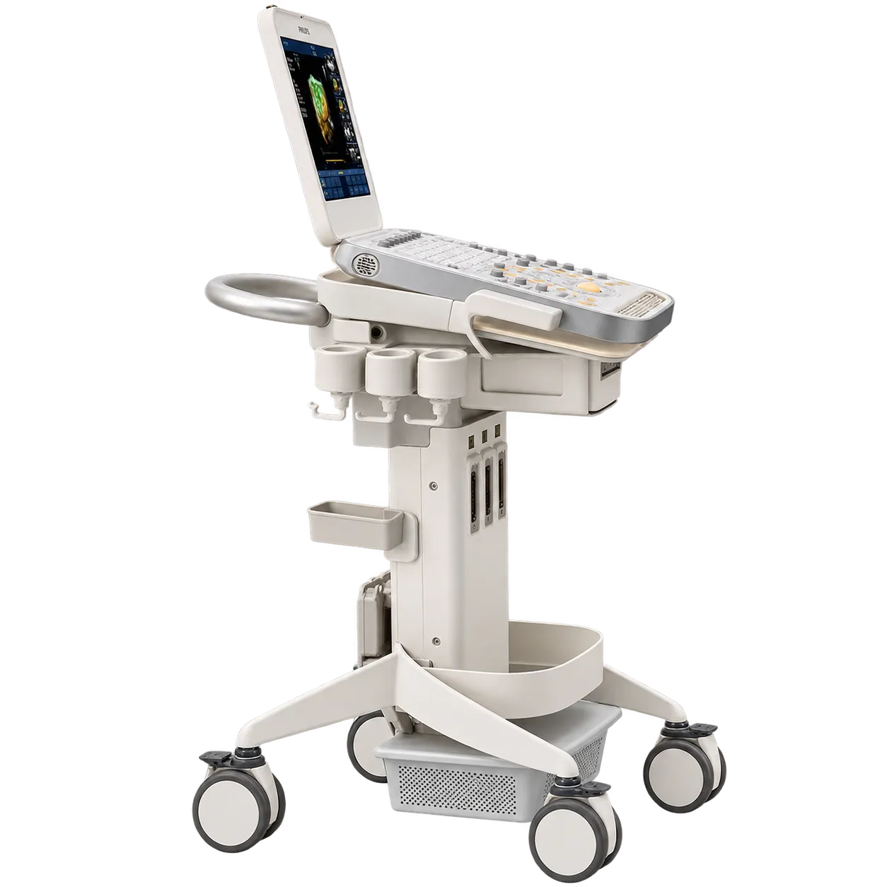
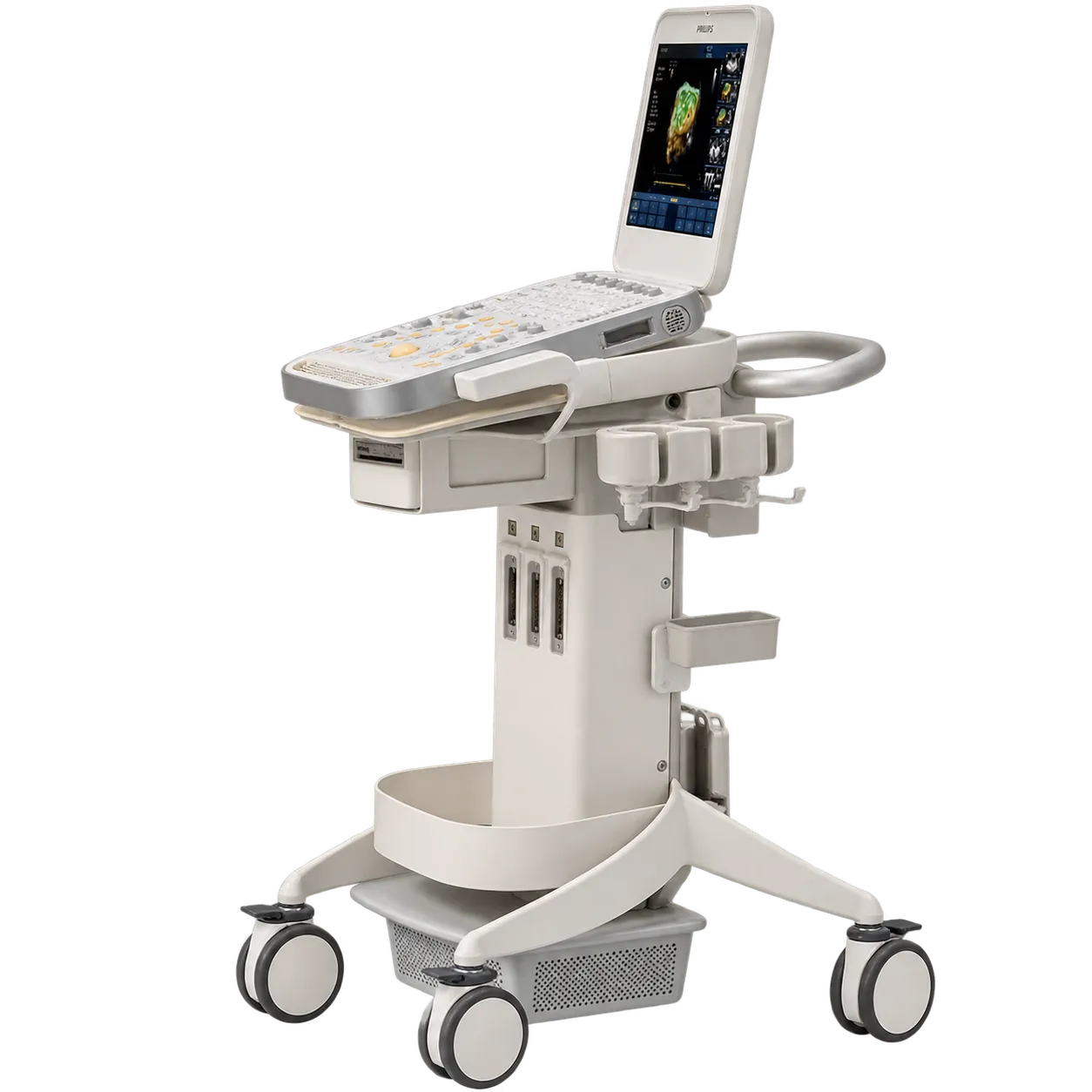
Vascular access is supported by high-frequency linear imaging, with vessel identification through compression behaviour and Doppler, and real-time visualisation of the needle as it advances. All clinical judgement, including whether a procedure is indicated and how it is performed, remains with the clinician and the service.
Diagnostic vascular assessment runs on the same transducer, covering vessel wall and lumen imaging with flow information.
Small parts work covering thyroid, testicular and other superficial structures uses the same linear probe with the relevant presets.
Superficial musculoskeletal imaging of tendons, muscles and joint structures is served by the same transducer.
Abdominal and cardiac work is covered where curved array and phased array transducers are among those supplied.
Critical care, theatre and treatment room use is supported by carrying the system to the location and working on internal battery.