
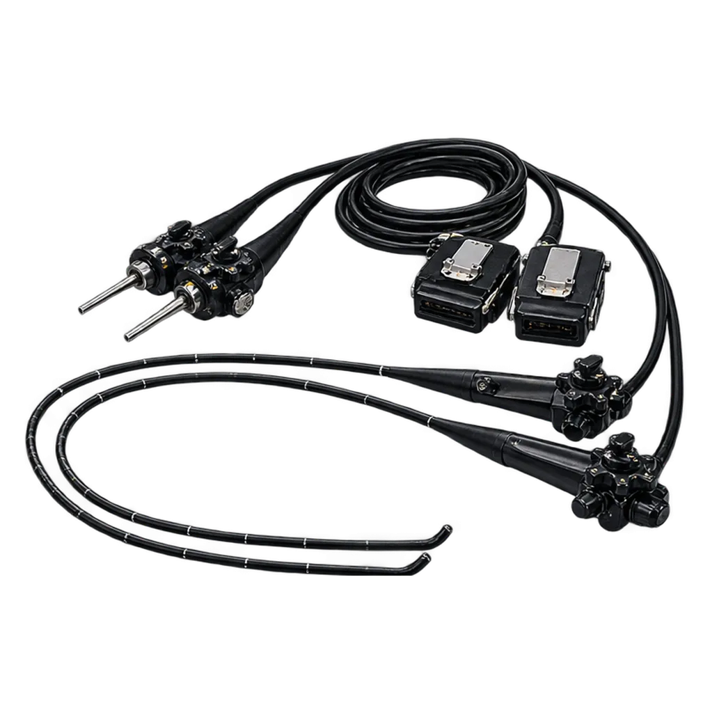
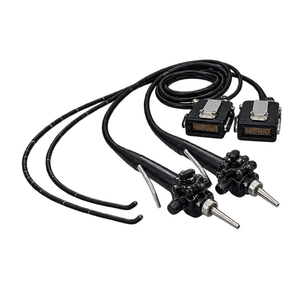
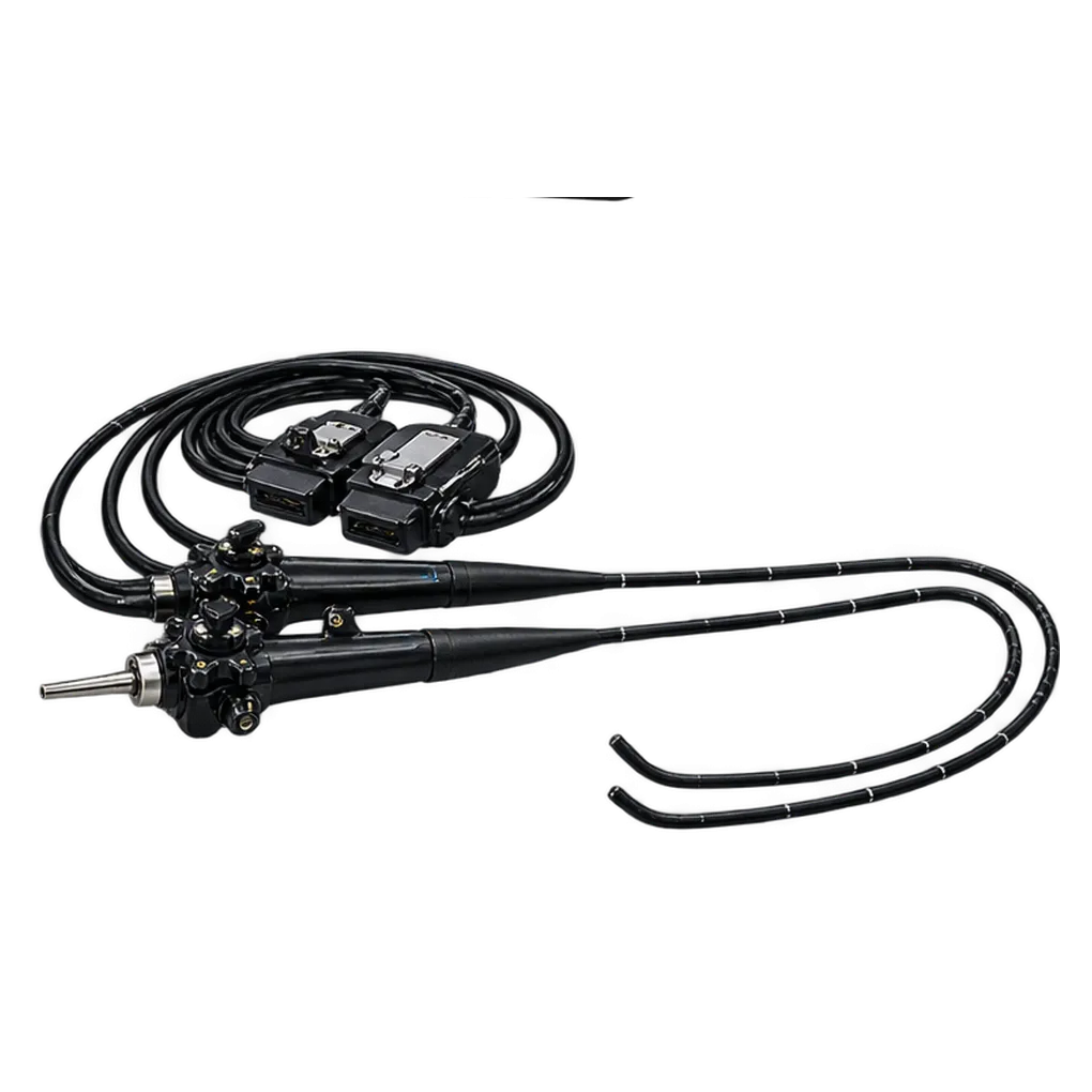
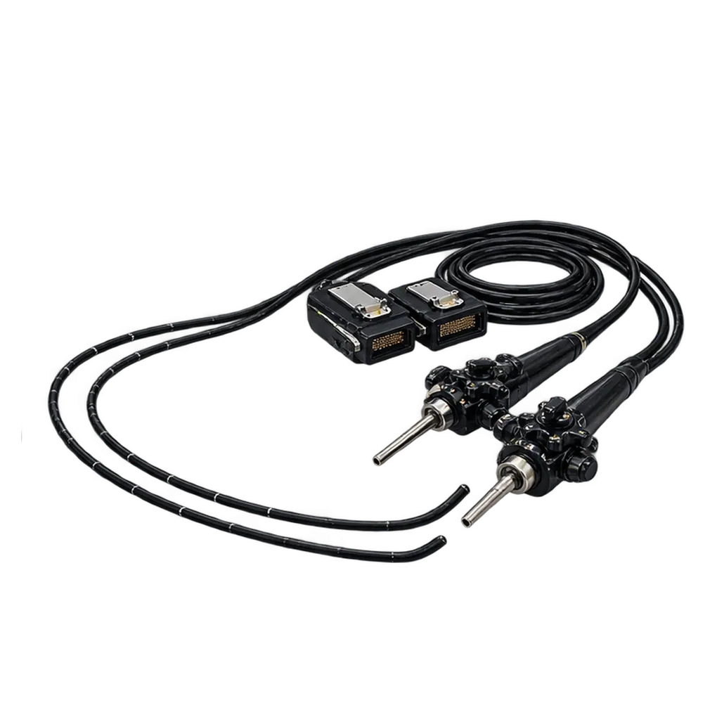
The GF-UCT160-OL5 belongs in endoscopy suites where endoscopic ultrasound forms part of the daily list. Hospital gastroenterology departments are the natural home for a linear echoendoscope, and the room holds the EU-C60 ultrasound centre beside the scope.
EUS-trained endoscopists operate the instrument. Assistants prepare the contact balloon or the sterile deaerated water and handle needles and accessories. Four-direction bending lets the operator steer the transducer face toward the tissue under examination, while the 55° oblique video view keeps the tip in sight during positioning.
The first use is imaging of the upper gastrointestinal tract and the structures beside it. The 150° sector at 7.5 MHz forms the B-mode image, and colour power Doppler adds blood-flow display. Acoustic contact comes from a balloon or from water immersion, and the operator chooses between them for each examination.
The second use is sampling. Once the scan has located a target, a 22G fine-needle aspiration needle passes through the 3.7 mm channel, so imaging and sampling share one insertion. That workflow suits units that run diagnostic and tissue-sampling sessions in the same room with the same equipment. A 100° video view and a depth of field from 3 to 100 mm support positioning before each scan.
A session follows the sequence built into the scope. The video view positions the tip, the balloon or the water establishes acoustic contact, and the 150° B-mode image shows the target. Colour power Doppler adds blood flow to the display, and a 22G needle then passes through the channel along the route the image has defined. Each stage runs through the same 12.6 mm insertion tube.