


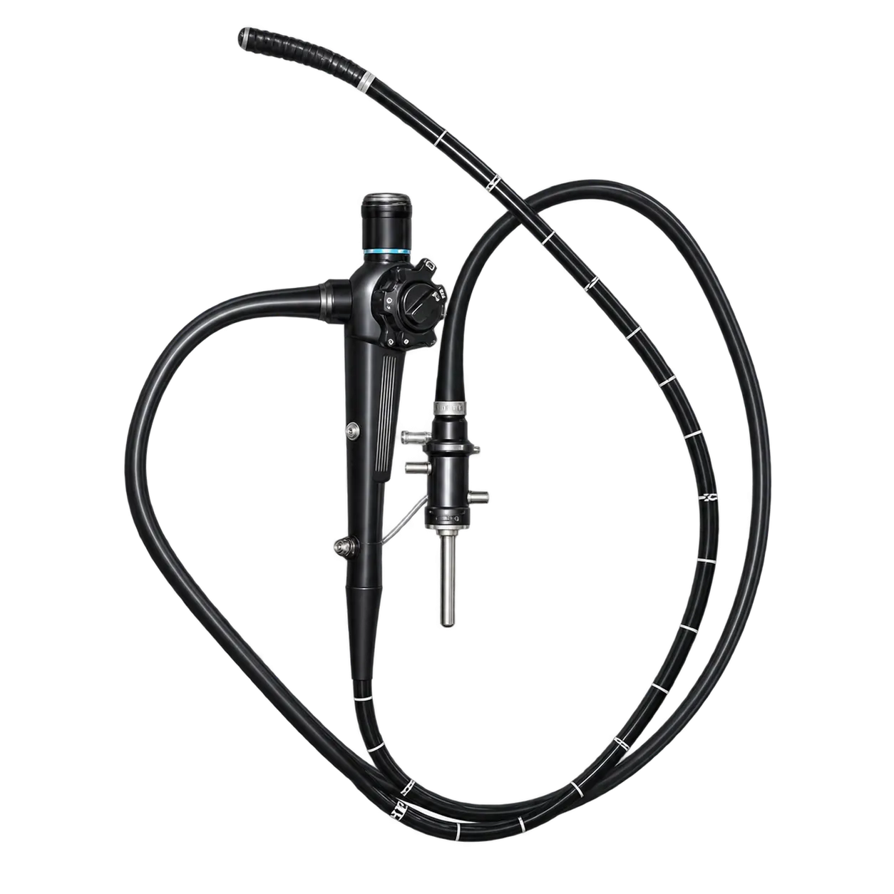
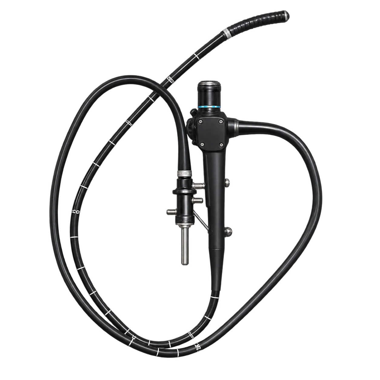
Clinical Applications
The GIF-1T20 serves endoscopy suites and gastroenterology departments that treat and examine conditions of the upper digestive tract. The endoscopist steers the scope and views the image through the eyepiece, while the assisting team prepares the light source and treatment instruments before each session.
In use, the scope travels from the oesophagus into the stomach and on to the duodenum. A 100° field of view captures a broad area of the lining, so the wall surface and the course of the passage appear together in one image.
Simplicity shapes the routine. Glass strands carry the image to the eyepiece, so the examination needs no video sensor, processor, or monitor. The team sets up the scope and its light source, and the endoscopist looks straight into the picture.
The 3.7 mm channel serves the treatment. Instruments sized for the bore pass down the channel to the target, so a single insertion carries both examination and treatment.
Four-way bending of 210° up, 90° down,n and 100° to each side lets the endoscopist aim the tip and the channel at each wall in turn. The 11.2 mm tube and 1,030 mm insertion length carry the tip to areas of interest in the upper tract.
Departments that keep a light source in the room can place the scope on a routine list at once. The endoscopist and the team then follow each step from the first look to the last treatment.