
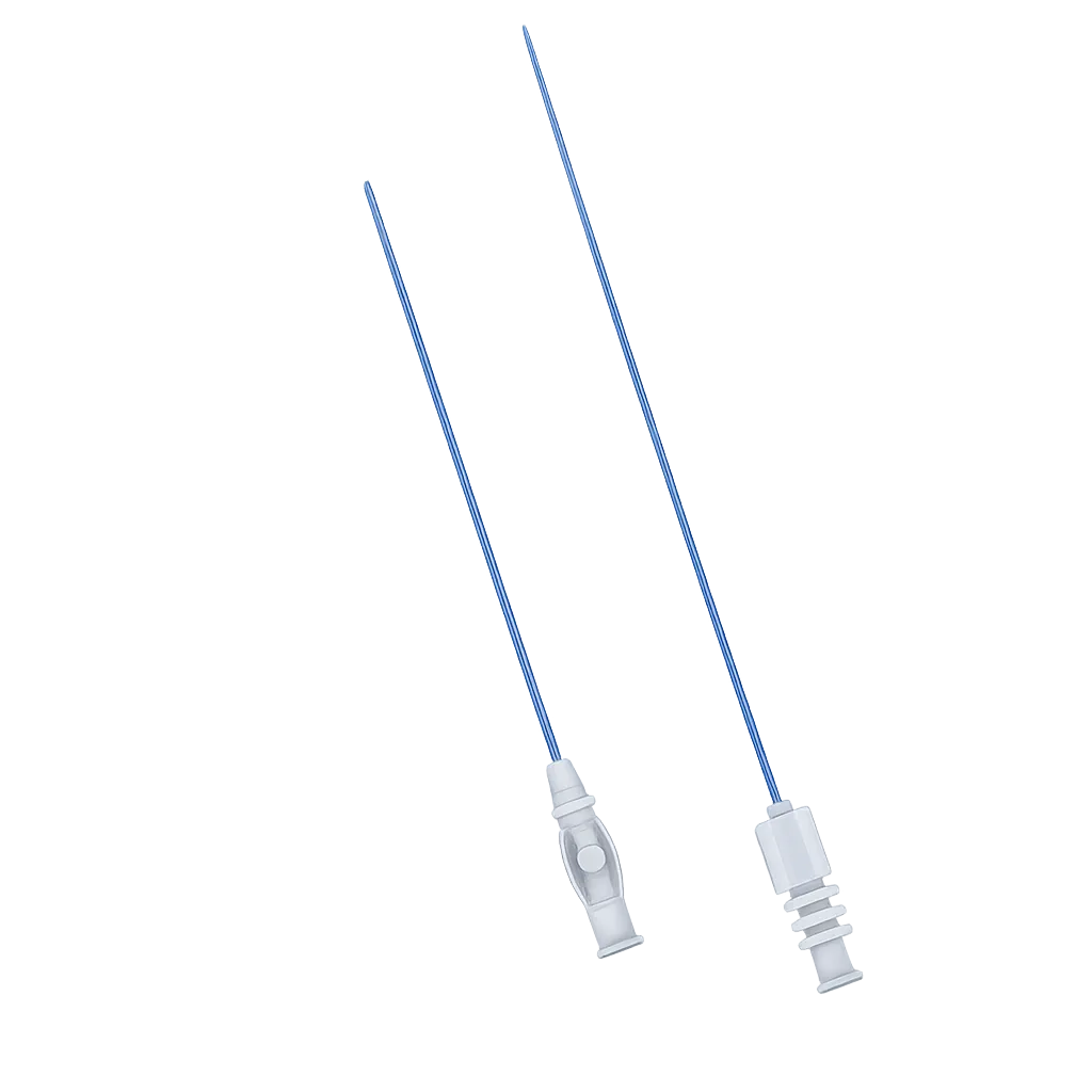
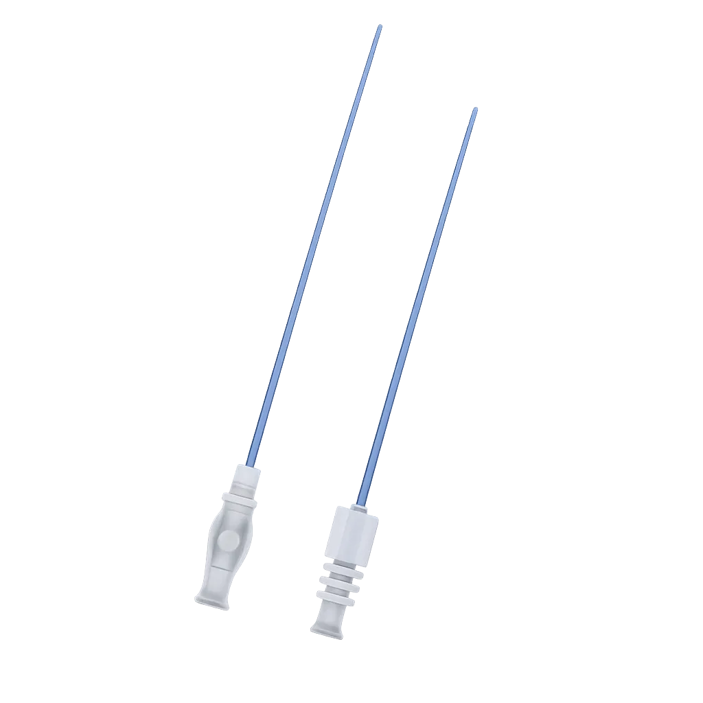
Vascular access usually begins with a needle passed blindly or under ultrasound into an artery or vein. The conventional approach uses an 18G needle, which is large enough to take a 0.038-inch wire directly. Micropuncture takes a different route, entering with a 21G needle and a much finer wire, then building the tract up in stages.
The argument is about what happens when a pass fails. An 18G needle that enters the back wall of a vessel, or crosses a branch, leaves a hole that bleeds. A 21G needle leaves a considerably smaller one. That matters most where the consequences of a poor pass are greatest, in patients on anticoagulation, in vessels that are calcified or difficult to feel, and in access sites where bleeding would be hard to control. The published evidence is not unanimous. One retrospective cohort of nearly eighteen thousand patients found significantly fewer groin haematomas and fewer complications overall with the fine needle, while a smaller comparison found no measurable advantage.
Ultrasound guidance is where the technique now sits most comfortably, and it is why the echogenic needle option matters. A fine needle is hard to see on ultrasound, especially when approaching at a steep angle, and a tip treated to scatter sound is far easier to follow. One caution belongs alongside all of this. The fine wire that follows the needle can be diverted into a small side branch more readily than a stiffer J-tipped wire, and perforation has been described.
Handling the wire gently and confirming its course remains part of the technique.Medigear.uk supplies the instructions for use with every order.