
Vertebral compression fractures collapse the front of a vertebral body, causing pain and a forward stoop. Kyphoplasty treats them in two stages. A balloon is inflated inside the bone to push the collapsed trabeculae apart and create a cavity, sometimes restoring some lost height. Cement is then placed into that cavity.
The balloon provides control. In vertebroplasty, cement is forced directly into intact cancellous bone, which resists it, and the pressure needed is exactly what drives cement into places it should not go. A prepared cavity accepts cement at lower pressure, which is the main argument for the extra step. Cement escaping the vertebral body remains the complication that matters most in both procedures, since it can compress the spinal cord or travel through veins to the lungs.
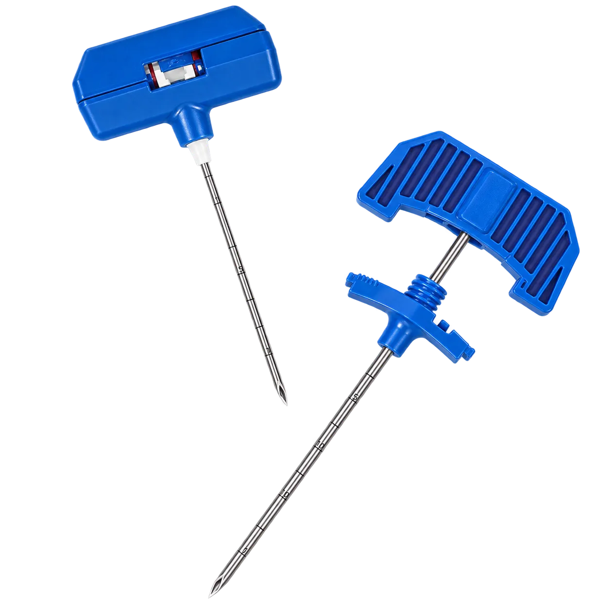
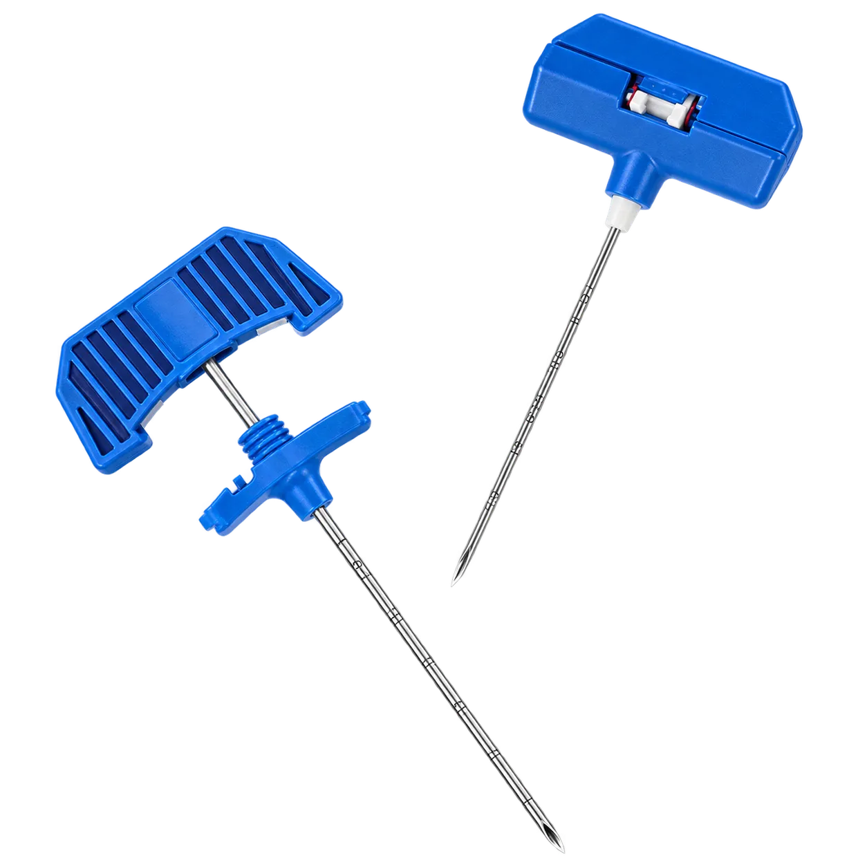
Everything in this set serves the first part of that sequence. The channel must be accurate, because it passes through the pedicle,e with the spinal canal immediately medial to it. A needle that strays through the pedicle wall can reach the dura, and cement following that track has a direct route to the cord. For this reason, imaging guidance is used throughout, and each instrument is exchanged over the previous one rather than creating a new track. That is why the guide wire matters, and why the dimensional fit between components is worth checking rather than assuming. In some cases, bilateral access through both pedicles is used, which doubles the instrument requirement for that level.
Medigear.uk supplies the instructions for use with every order.