
Infection in a joint replacement is among the hardest problems in orthopaedics. Bacteria form a biofilm on the implant surface. Antibiotics given by drip struggle to penetrate it. Reaching an effective concentration in the joint that way would mean doses the kidneys could not tolerate. Placing antibiotic directly into the cavity solves both problems at once.
Beads are how that is usually done. A carrier material is loaded with antibiotic, formed into beads, and left in the surgical site. Local concentrations many times above the level needed to kill the organism are achievable. Blood levels stay low. The carrier determines much of what follows. PMMA cement releases antibiotic from its surface rather than throughout. Concentrations are lower and sustained release is limited. It does not dissolve either, so the beads must usually be retrieved at a later operation. Calcium sulfate carriers dissolve instead, releasing antibiotic more completely and leaving nothing to remove.
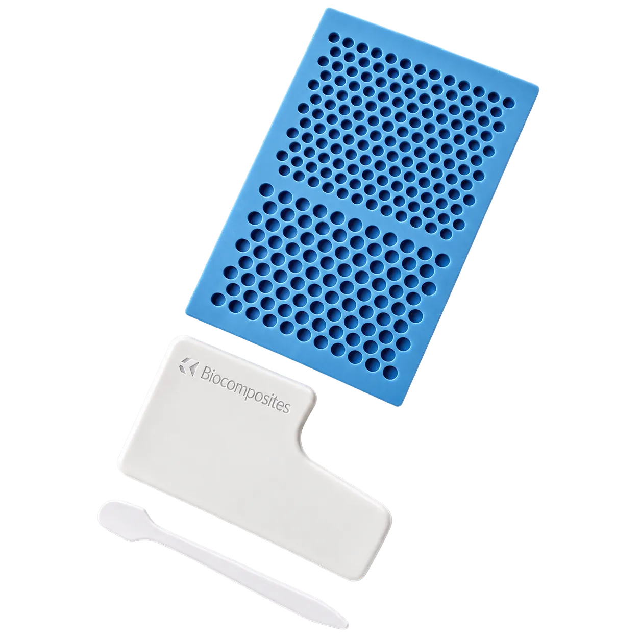
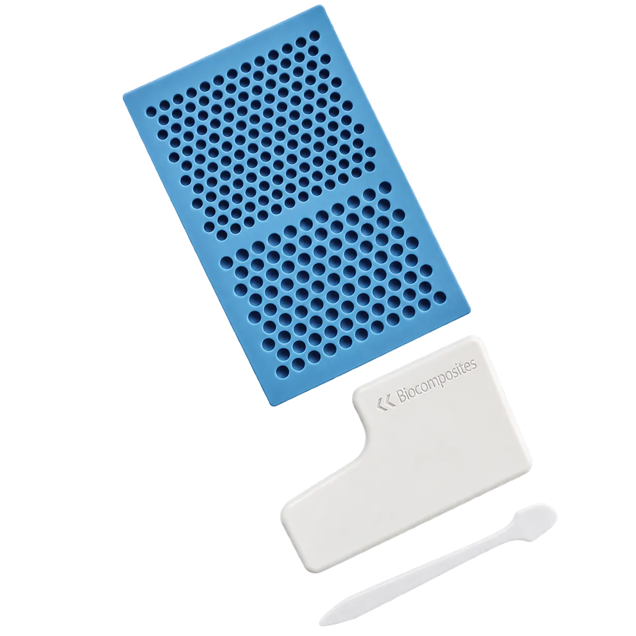
Bead size is the variable the surgeon controls. Smaller beads have more surface area relative to their volume. They release faster, which suits shorter courses or a high early dose. Larger beads release more slowly and fill space more efficiently. Published work in this area has found smaller beads eluting faster. Combining two antibiotics has been shown to extend release considerably. A mat offering three sizes allows both in the same procedure. Larger beads pack the bulk of a cavity while smaller ones deliver a faster initial dose.
Medigear.uk will supply the instructions for use once the documentation is complete.