
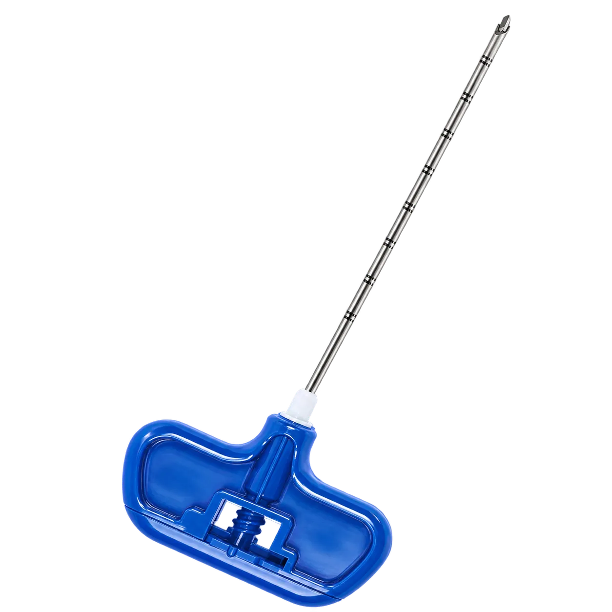
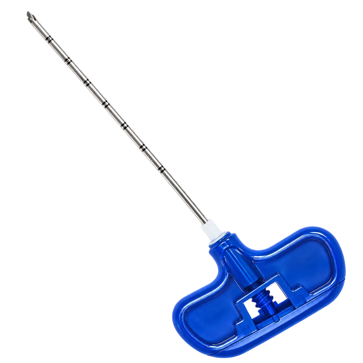
Bone lesions seen on imaging often cannot be characterised from the images alone. A sclerotic patch, a lytic hole or an area of marrow signal change may represent a metastasis, a primary tumour, infection or something benign, and treatment differs entirely between them. Taking tissue settles the question.
For most of these questions, the sample has to be a core rather than an aspirate. Aspiration collects cells, which suits cytology and culture. Histology needs the tissue held in its original arrangement, because how cells sit relative to one another distinguishes many bone lesions. A trephine cuts and captures that arrangement intact, which is why toothed cutting needles rather than plain ones are used for bone.
Two technical problems limit diagnostic yield, and both concern the sample rather than the targeting. The first is inadequate size, where too little tissue is retrieved for a pathologist to commit to a diagnosis. An 11-gauge core is generally enough that one pass suffices, and can often be divided between histology and microbiology. The second is crush artefact, where the sample is compressed during cutting or extraction and the architecture that made it worth taking is destroyed. Steady rotation under moderate pressure addresses the first part, and gentle expulsion with the probe addresses the second. Vertebral lesions are usually approached through the pedicle under CT or fluoroscopic guidance. Sampling more than one part of a large lesion is common, since the diagnostic tissue may not be evenly distributed within it.
Medigear.uk supplies the instructions for use with every order.