
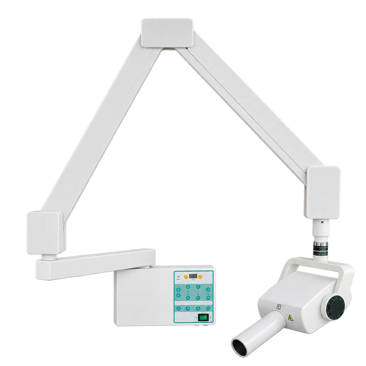
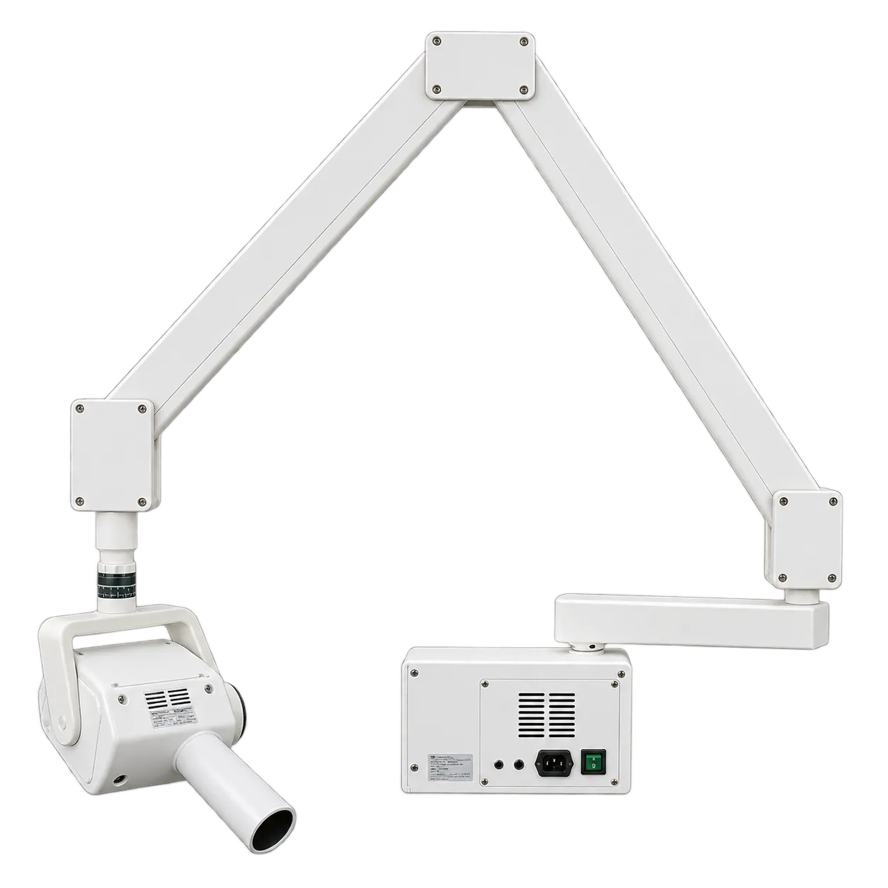
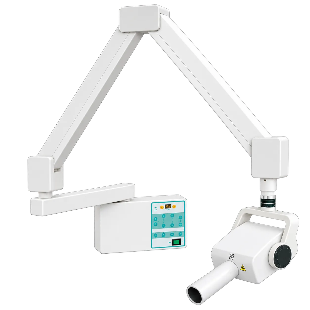
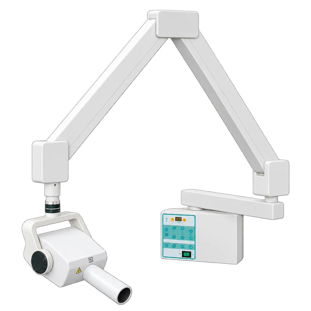
General dental practices fit the HS-X101 at the chair where intraoral radiographs are taken most often, so periapical and bitewing images are taken without the patient moving. The articulated arm brings the tubehead to the cheek for each angle, and folding it back to the wall returns the space around the chair to the dental team.
Endodontic practices take working-length and post-obturation radiographs within one appointment, while implant teams image each fixture after placement and again at review. Practices that fit one unit per surgery give every clinician an X-ray within reach, with the same light-touch controls in each room. Community dental services and multi-surgery groups standardise on one wall unit per surgery, so a locum finds identical controls in every room. Orthodontic and paediatric lists use it for bitewing checks at routine reviews.
Dental nurses prepare the sensor, film or phosphor plate and the holder, while the clinician sets the exposure time on the control panel. Teams moving from film to digital keep the same unit, since it works with intraoral sensor systems and film alike. The optional 0.5 mA, 0.8 mm configuration suits practices whose work leans on fine detail, such as endodontic files and root outlines.
Training clinics and dental school chairs suit a fixed unit as well, because every student meets the same control panel and the same arm movement at each chair.
It does not suit domiciliary visits or imaging spread across several sites, which call for a portable source. Panoramic and cephalometric imaging need an extraoral system, and the stated supply is 220 V ± 10% at 50 Hz.