
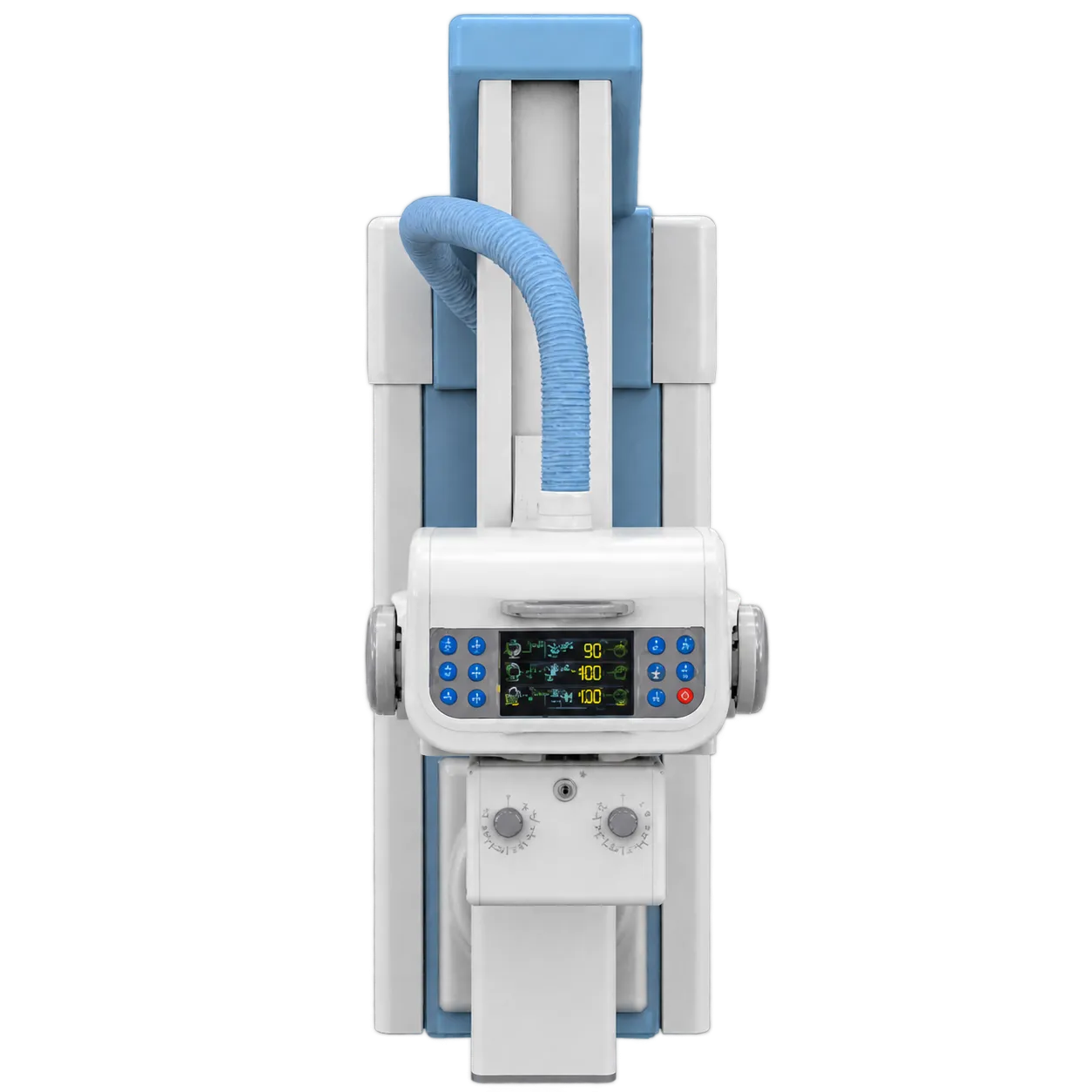
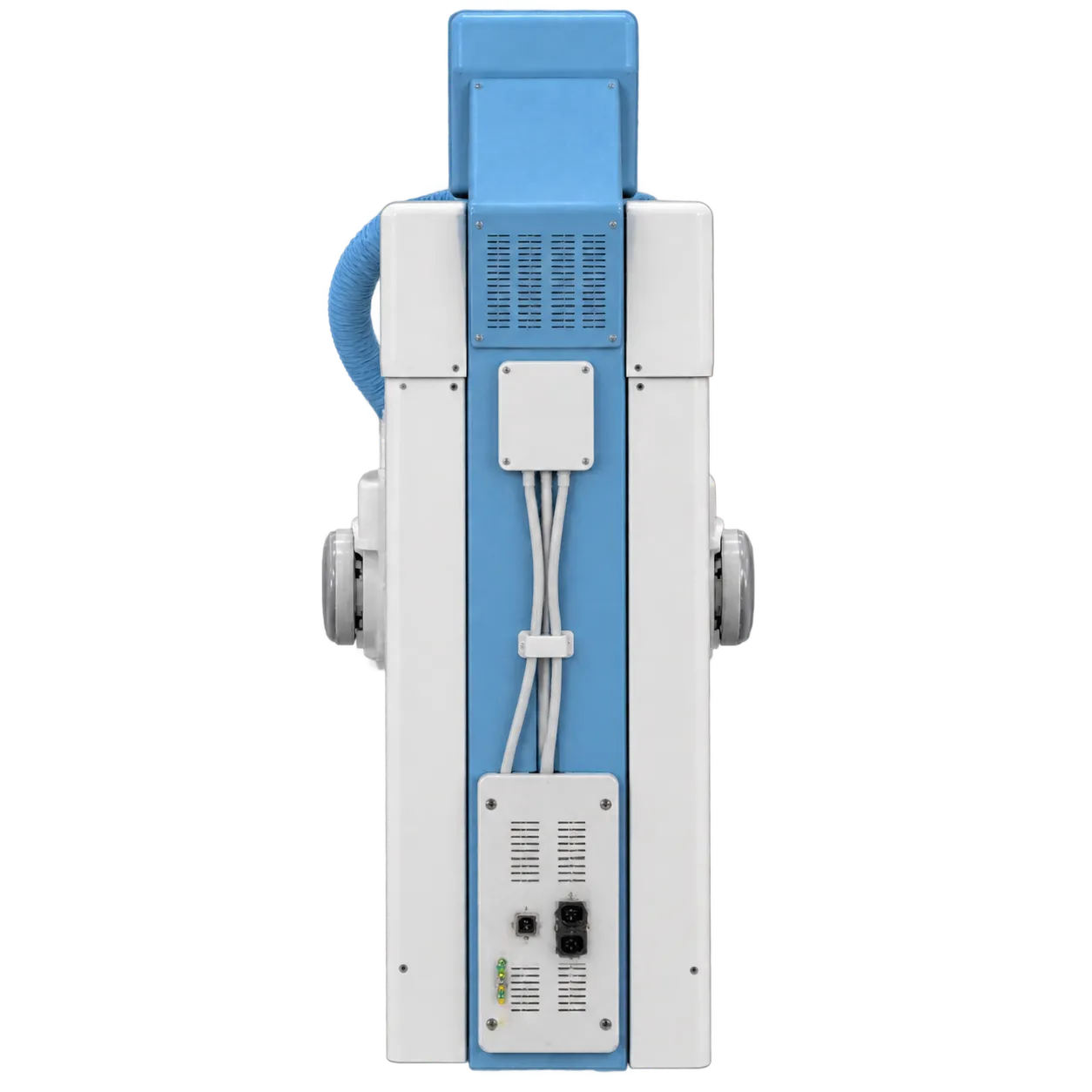
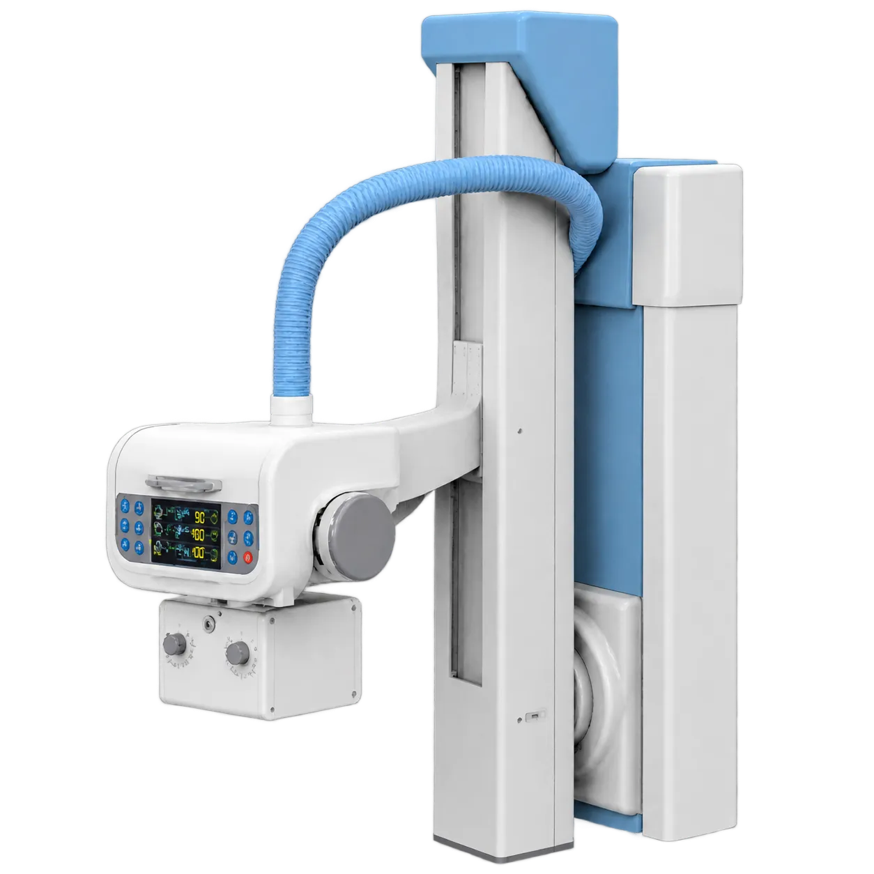
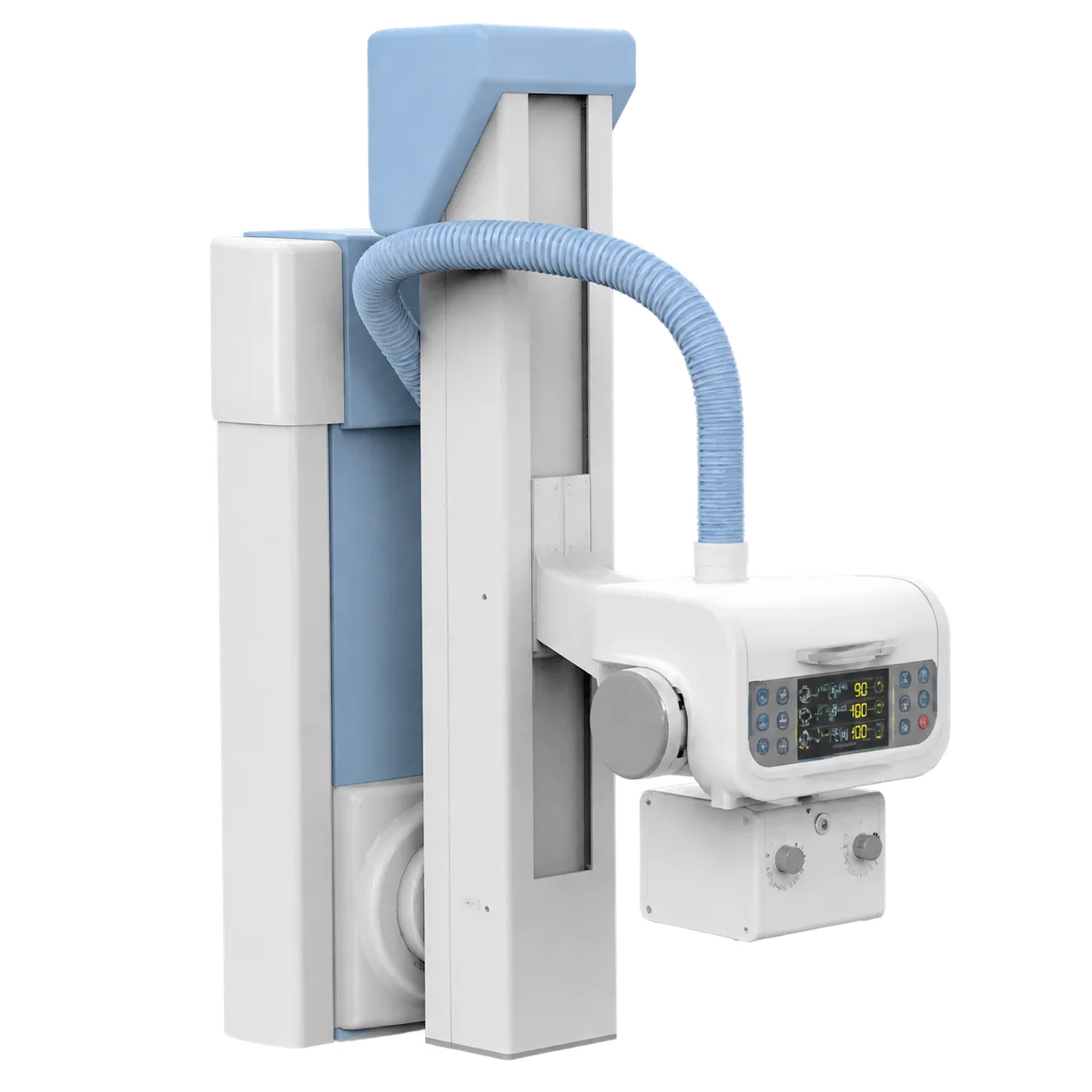
Collimation is the simplest dose control in radiography. Restricting the beam to the area under examination cuts the volume of tissue irradiated. It also reduces scatter and improves contrast, and UK practice treats it as routine for every exposure. A manual X-ray collimator puts that entirely in the radiographer's hands. With good technique and a working light beam, it is perfectly serviceable, and rather less forgiving without one.
The light itself therefore matters more than the sheet suggests. Staff line the field up by what they can see, so the lamp needs to be bright enough in a lit room. The light field also has to agree with the radiation field. Physics teams check that agreement at commissioning and at intervals afterwards, along with the accuracy of the field size indicators on the control face. A timer saves the bulb, and spare bulb availability is worth asking about at the outset, since a dark collimator stops careful work.
Filtration sits in the same category of quiet essentials. It removes low-energy photons that would be absorbed in the patient without ever reaching the detector;d those photons contribute dose while adding nothing to the image. The total for the assembly therefore has to be established and recorded, and that figure belongs in the room's documentation alongside leakage radiation, which is also absent here.
Medigear.uk obtains the leakage data, field size coverage, and mounting details from Blackstone, so the radiation protection adviser can complete the assessment without chasing the manufacturer later.