
Laparoscopic and General Surgery
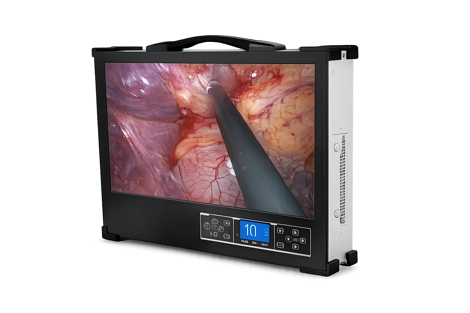
Abdominal work is where three-sensor capture earns its place, because the surgeon separates peritoneum, fat and vessel by hue as much as by contour. Dedicated sensors report those distinctions honestly instead of inferring them, and the 1300 line vertical format keeps a circular scope view generously presented. Integrated illumination at 120 watts reaches the depth abdominal work requires while keeping the whole arrangement to a single unit on the bench.
Gynaecology and Urology
Examination through confined passages benefits from accurate colour, since mucosal appearance carries diagnostic weight and a colour cast can quietly mislead. Three-sensor separation holds those tones faithfully throughout a session. The integrated display suits clinic rooms where no monitor was ever mounted, and retained VGA connects readily to whatever screen a room already happens to have installed. Stored material remains retrievable once the clinic session has finished.
Otolaryngology and Orthopaedics
Airway, sinus and joint work all involve confined reflective spaces where tissue types share similar tones and separation matters. Faithful colour helps distinguish surface texture and early change that flatter rendering would obscure. The portable format suits rooms serving several functions, since the unit arrives complete and leaves when the session ends. Nothing has to be mounted or wired into the room beforehand.
Teaching and Case Documentation
Accurate colour makes recorded material genuinely useful for teaching, because trainees learn to recognise tissue by appearance and a distorted palette teaches the wrong lesson. USB transfer moves captured material onward for review and assessment, while the range of video outputs lets a teaching screen run alongside the built-in display during a live session.