
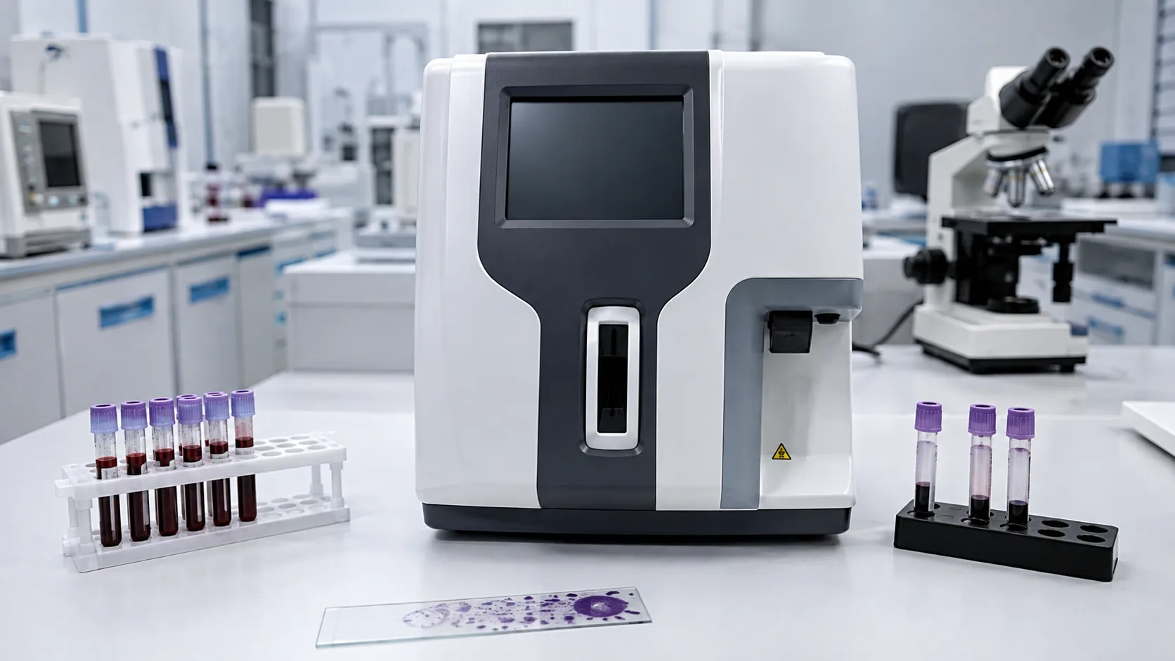
Reticulocyte counting measures the proportion and number of the youngest red cells in circulation, giving a direct read on how actively the bone marrow is making red cells. Reticulocytes still carry residual RNA, and analysers detect that RNA to separate them from mature red cells, reporting a percentage, an absolute count and often maturity indices. For a haematology buyer, the questions are which counting technology the platform uses, which indices it reports, and how the reticulocyte channel sits within the wider full-blood-count workflow.
The count helps distinguish causes of anaemia and shows the marrow's response to treatment. The analyser produces the figures; clinical interpretation belongs to the requesting team.
What a reticulocyte is and why it is counted
A reticulocyte is a red cell that has just left the bone marrow and has not yet fully matured. For roughly a day, it still contains ribosomal RNA, which precipitates into a mesh when stained. Counting these cells shows whether the marrow is responding appropriately, for example rising after blood loss or with effective treatment, or staying low when production is impaired. Because the window is short, the reticulocyte count is a near-real-time indicator of marrow activity, which is why it is used to monitor response rather than to make a single diagnosis.
How reticulocyte counting devices work
Modern analysers count reticulocytes by flow cytometry. A fluorescent dye binds the residual RNA, cells pass single file through a laser beam, and the instrument measures scattered light and fluorescence for each cell. The fluorescence intensity reflects RNA content, so the analyser both identifies reticulocytes and grades their maturity, reporting low, medium and high fluorescence fractions. From these it derives the reticulocyte percentage, the absolute reticulocyte count and, on many platforms, the immature reticulocyte fraction and the reticulocyte haemoglobin content. Automated counting examines tens of thousands of cells, giving far better precision than the old manual method of counting supravitally stained cells under a microscope.
The indices a buyer should look for
Beyond the basic percentage and absolute count, several derived measures add clinical value and are worth confirming a platform reports. The immature reticulocyte fraction flags the very youngest cells and can signal early marrow recovery. The reticulocyte haemoglobin content reflects the iron available for recent red-cell production and can support assessment of iron status. Check which of these indices the analyser provides, how they are named on its reports, and whether the reference intervals are configurable, because different laboratories and clinical services rely on different subsets of these measures.
Integration with the full blood count
Reticulocyte counting rarely stands alone. On most modern haematology analysers it is one channel within a platform that also produces the full blood count and differential, so a single aspirated sample yields the reticulocyte data alongside haemoglobin, red-cell indices and white-cell counts. This integration matters for procurement: buying a reticulocyte capability as part of the main analyser avoids a separate instrument, shares reagents and sampling, and keeps everything on one result set. Confirm whether reticulocyte counting is standard or a licensed add-on, whether it runs on every sample or on request, and how it affects throughput when enabled.
Key specifications a buyer must check
Review the sample type and volume, whether the analyser samples from a closed EDTA tube, and the throughput in samples per hour with the reticulocyte channel active. Check the measuring range and precision for the reticulocyte count, the list of maturity and haemoglobin indices reported, and the reagent format and onboard stability. Assess the autosampler capacity, cap-piercing for staff safety, and how the platform flags interferences that can affect the count, such as nucleated red cells or certain inclusions. Confirm that reference intervals can be set for adult and paediatric populations and that the analyser links to the laboratory information system.
Regulation and applicable standards
A reticulocyte analyser is an in-vitro diagnostic device and must carry UKCA or CE marking backed by a declaration of conformity, with the manufacturer registered for Great Britain through the MHRA. Electrical safety is addressed by IEC 61010 and electromagnetic compatibility by IEC 61326, while a laboratory running the platform works under ISO 15189 for medical laboratory quality. The International Council for Standardisation in Haematology has published guidance on reticulocyte counting that underpins method comparison. General device procurement guidance for the public sector sits on GOV.UK. Verify the paperwork rather than accepting a specification sheet at face value.
Calibration, quality control and maintenance
Reproducible reticulocyte results depend on disciplined control. Run dedicated reticulocyte control materials at the intervals the manufacturer sets and after each service, record the values, and investigate any drift before it reaches reporting. Because the channel relies on a fluorescent stain and optical detection, keep the flow path clean to avoid carry-over, replace lamps or lasers and tubing on schedule, and follow the maker's routine cleaning cycles. Enrol in an external quality assessment scheme that covers reticulocytes so performance is benchmarked against peer laboratories. Keep control and maintenance logs for accreditation, and treat controls, reagents and periodic servicing as recurring budget lines.
Where reticulocyte counting is used
The count supports several services, and the platform should match the busiest of them. General haematology laboratories run reticulocytes as part of anaemia investigation and to track response to treatment. Renal and oncology services use the count and reticulocyte haemoglobin content to monitor red-cell production during therapy. Transfusion and transplant teams watch the immature reticulocyte fraction as an early sign of marrow recovery. A high-volume laboratory needs a large autosampler and the reticulocyte channel enabled across the workload, whereas a smaller site may run it selectively; either way, define who reviews flagged results and how they reach the record.
Consumables, connectivity and total cost of ownership
The reticulocyte reagent, stain and controls are the recurring costs, and they should be modelled at your real workload rather than a headline per-test figure. Confirm whether reticulocyte counting is bundled with the main analyser reagents or priced separately, and check onboard reagent stability to avoid waste on a lower-volume bench. Interfacing to the laboratory information system and any middleware should be verified so counts and indices post without transcription. Ask each supplier about warranty terms, first-service cover, loan cover and the cost of failure-prone optical parts. Buying the capability on an analyser you already run usually gives the lowest total cost; the MediGear buyers team can help compare options, or contact us for availability.
Procurement checklist
-
Confirm the counting technology and that flow-cytometric fluorescence detection is used.
-
Check which indices are reported, including immature fraction and reticulocyte haemoglobin.
-
Decide whether reticulocytes run on every sample or on request, and the throughput impact.
-
Verify closed EDTA-tube sampling, autosampler capacity and cap-piercing.
-
Confirm UKCA or CE marking, IEC 61010 and MHRA registration on paper.
-
Set reticulocyte controls, maintenance schedule and external quality assessment.
-
Check that reference intervals are configurable for adult and paediatric use.
-
Model reagent and control cost at your workload and confirm laboratory-system interfacing.
Conclusion
Reticulocyte counting gives a fast, quantitative view of bone-marrow activity, and modern flow-cytometric analysers deliver it precisely alongside the full blood count. Focus procurement on the indices the platform reports, how the channel fits your workflow, and the discipline of controls and quality assessment behind every result. Comparing verified specifications through MediGear helps your laboratory choose a reticulocyte capability that supports reliable monitoring for the clinical teams it serves.
Disclaimer
This article is for informational purposes only. It is published by MediGear (medigear.uk) for general information and procurement guidance, and is not clinical, diagnostic, treatment, technical, engineering, legal or regulatory advice, nor a product endorsement, guarantee or substitute for professional assessment. MediGear does not provide medical consultations. Buyers should consult their clinical, biomedical, estates and regulatory contacts, and the manufacturer's documentation, and independently verify all specifications, certifications, compatibility and suitability before purchase. Specifications, certifications and availability are correct at the time of publication and may change without notice. MediGear is a medical-equipment distributor and does not sell medicines or pharmaceutical products.