
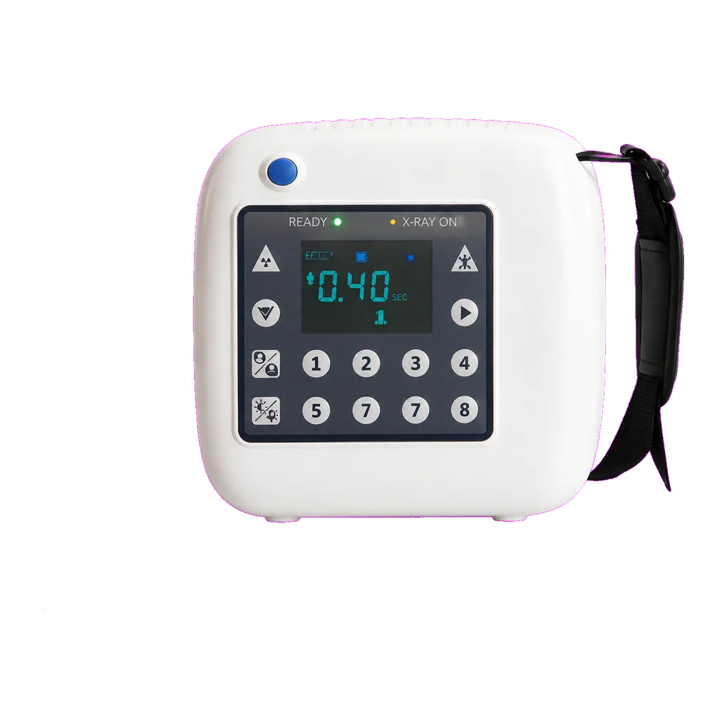
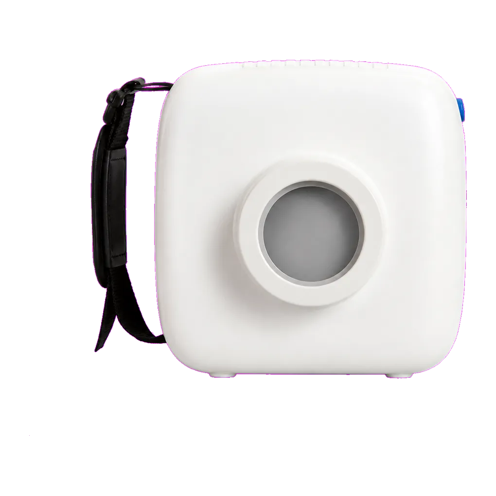
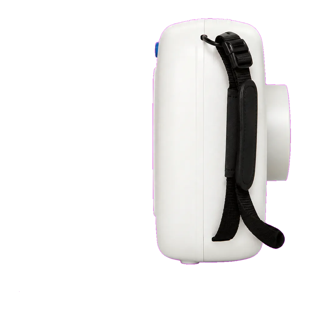
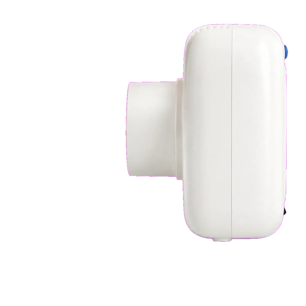
Intraoral radiography carries the diagnostic load in UK general dentistry, and the SJD-312 is configured for that daily volume. It supports bitewing caries detection, periapical assessment of pathology, endodontic length verification, alveolar bone-level review and root morphology assessment ahead of extraction.
Denture and edentulous care is where this unit is positioned, and it is a large, under-equipped population. Retained roots left at extraction are common and often silent until a denture presses on one. Buried teeth, sharp bony spicules and radiolucencies in healed extraction sites all show on periapical views, and any of them can explain a denture that will not settle or a sore spot that keeps recurring. A residual cyst — one left behind after the associated tooth was removed — is frequently found this way rather than sought.
Special care dentistry and community dental services carry much of that caseload. Older people in care homes, people living with advanced dementia, and patients receiving palliative care often cannot transfer to a fixed dental chair, so assessment happens at the bedside or in a day room. A carried unit is what makes the radiograph possible at all, provided off-site radiation protection arrangements have been agreed with the practice's Radiation Protection Adviser in advance.
Technique deserves specific attention with this population. Receptor holders are designed to be stabilised by the patient biting on them, and an edentulous ridge removes that anchor, so occlusal views, modified holder technique and cotton wool packing become the working methods. Operators should be trained in them explicitly.
Prosthetic planning uses these views for localised questions, alongside a panoramic radiograph where a whole-arch view of ridge form and sinus position is what the clinician actually needs.
Guidance from the College of General Dentistry and UKHSA Dental X-ray Protection Services remains the reference point UK practices work to, alongside HSE enforcement of IRR17.