
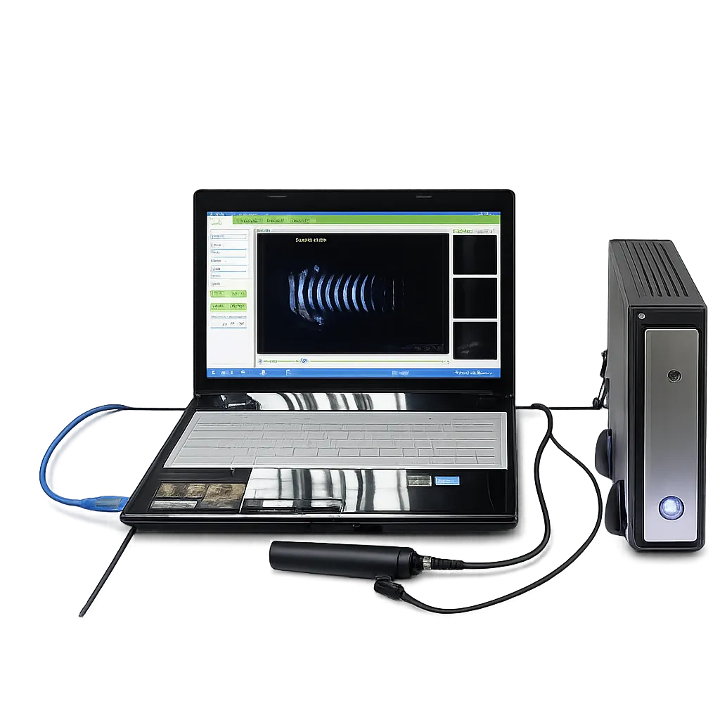
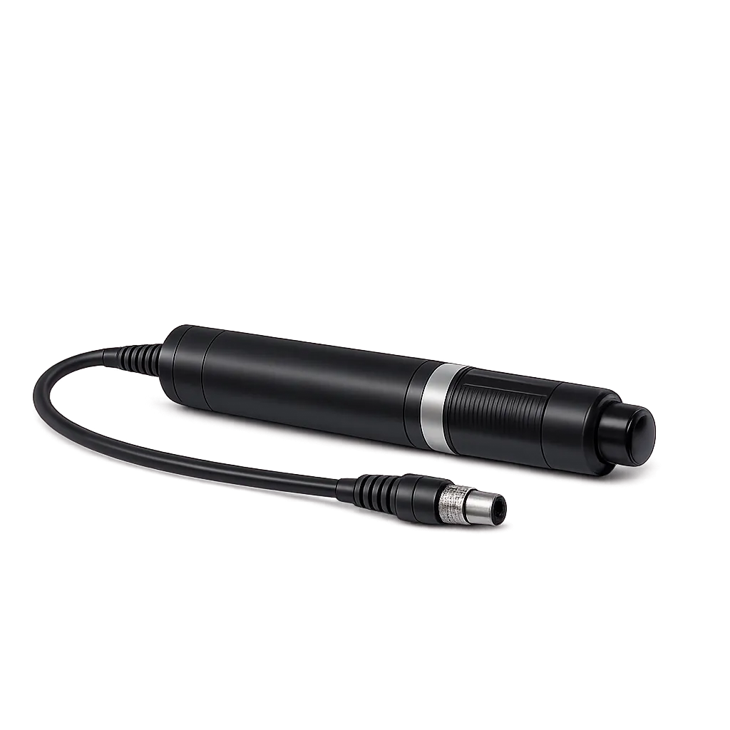
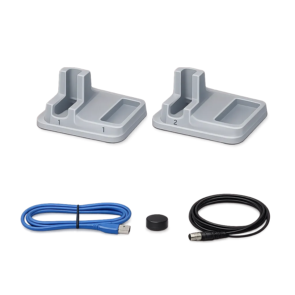
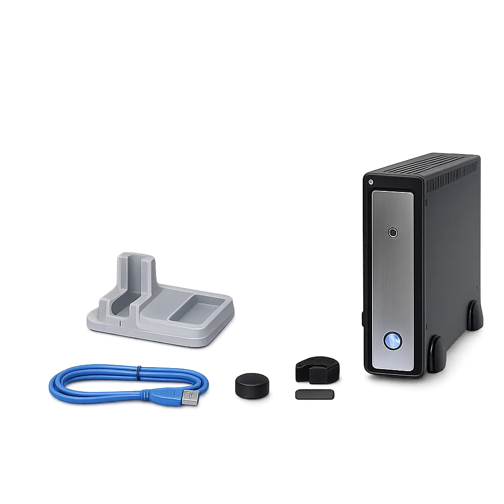
Cataract and IOL planning services use the A-scan side, which operates at 10 MHz to a depth of 40 mm and measures anterior chamber depth, lens thickness, and vitreous body length across phakic, aphakic, dense, and various IOL eye modes. Six formulae are listed,, including SRK-II, SRK-T, Hoffer-Q, Hollada,y and Binkhorst-II, a range that tracks axial length across short, averag,e and long eyes. Medigear.uk confirms the complete formula set and adjustable constants in writing before any procurement decision, since those determine whether the system fits an existing surgical protocol.
Ophthalmology clinics use B-scan where the fundus cannot be viewed directly. The manufacturer stathat tes the system is used the for diagnosis of intraocular diseaincludingming vitreous opacity, retinal detach,ment and ocular fundus tumours, with normal, vitreous body enhance,ment and retina observation modes provided. Those are the manufacturer's stated applications, and all interpretation remains with the practitioner. Detailed B-scan parameters are obtained from the manufacturer rather than assumed from related models.
Services with established IT arrangements may prefer a PC-based system, since data sits on practice hardware under practice access control and backup rather than on an instrument. That is genuinely an advantage where policies and systems already exist. It becomes a liability where the software requires an operating system the organisation cannot support, which is why Medigear.uk settles the platform question first.
Practices weighing entry cost against integrated hardware should note that a PC-based system defers the display and computer cost rather than removing it. MedigearUKk provides the computer requirements sothat the total cost can be assessed properly.