
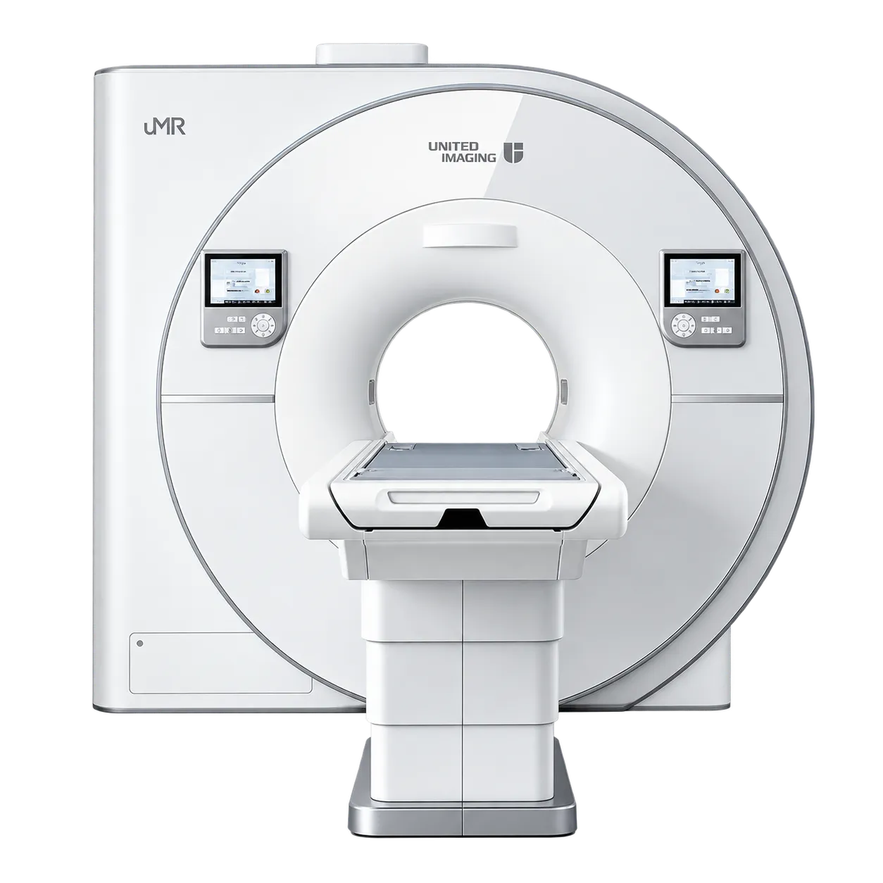
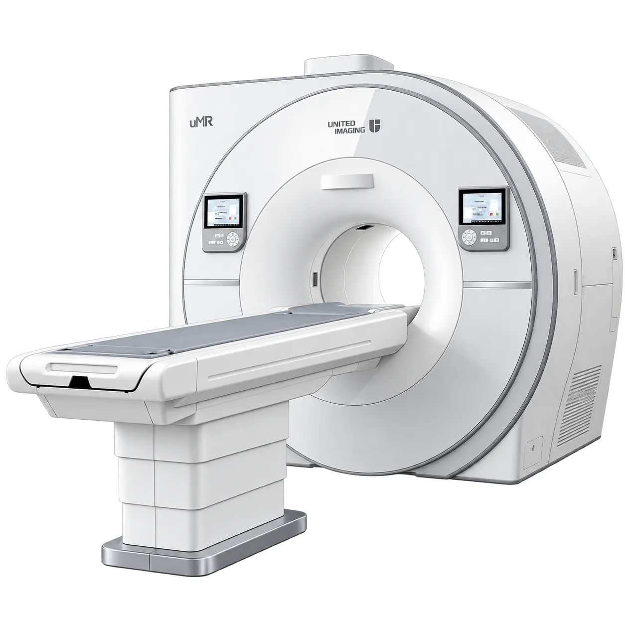
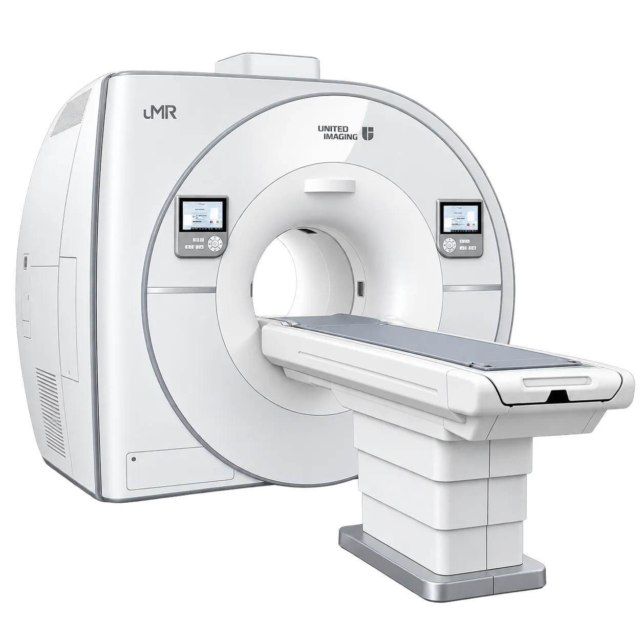
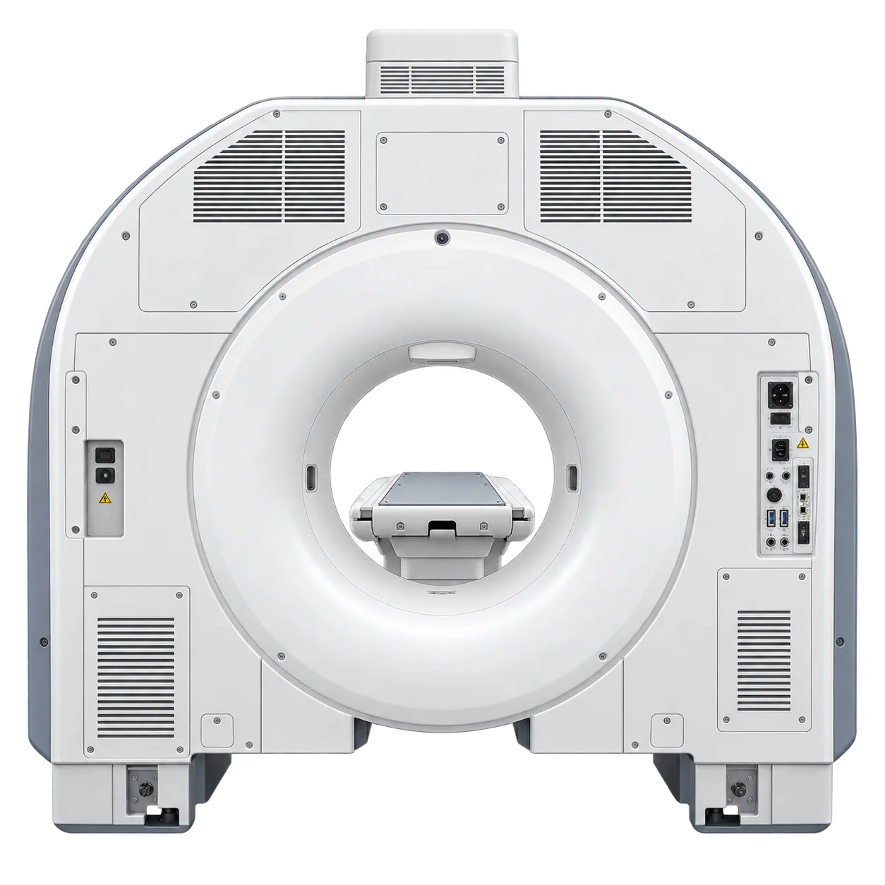
Neurological imaging benefits directly from acceleration, since long sequences can be shortened or resolution raised within the same acquisition window.
Musculoskeletal imaging draws on sub-millimetre isotropic acquisition, which allows a single dataset to be reformatted along the plane a joint requires.
Body and abdominal imaging is supported by field homogeneity across a large field of view, which underpins spectrally selective fat saturation.
Cardiac imaging depends on acceleration to bring breath-hold sequences within reach, alongside the gradient performance supplied on the individual system.
Off-centre imaging is supported by the short-magnet design and shimming, which holds homogeneity away from isocentre.
Patients at the upper end of size are accommodated by a 65 cm bore and a table taking 250 kg, which at 3 tesla is often the limiting factor on who can be scanned.
Departments under pressure on capacity are served by acceleration taken as shorter examinations, since throughput follows directly from sequence length.