
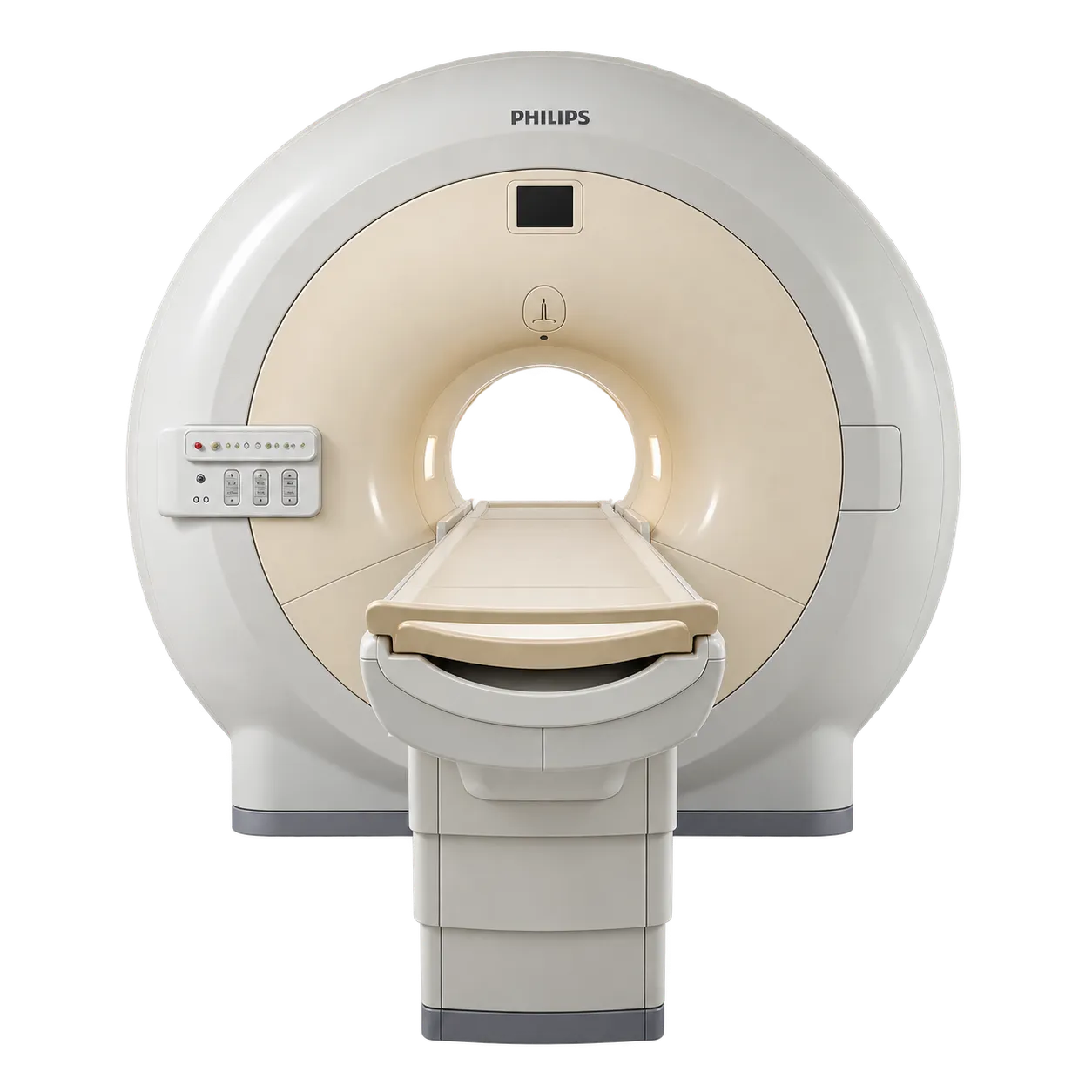
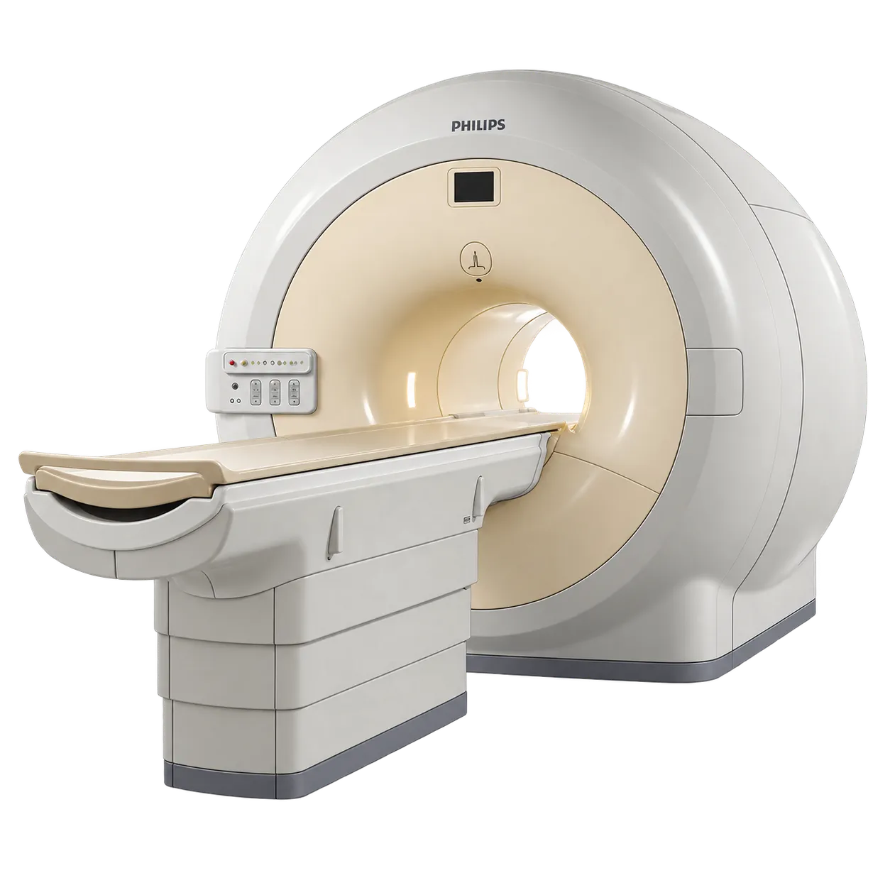
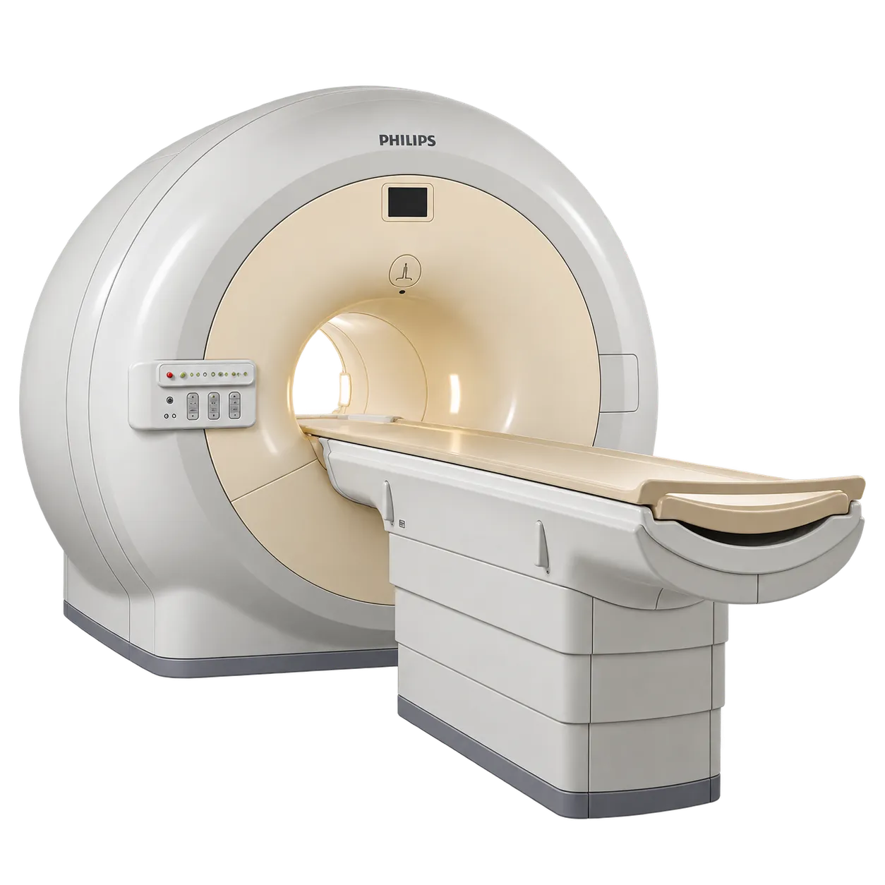
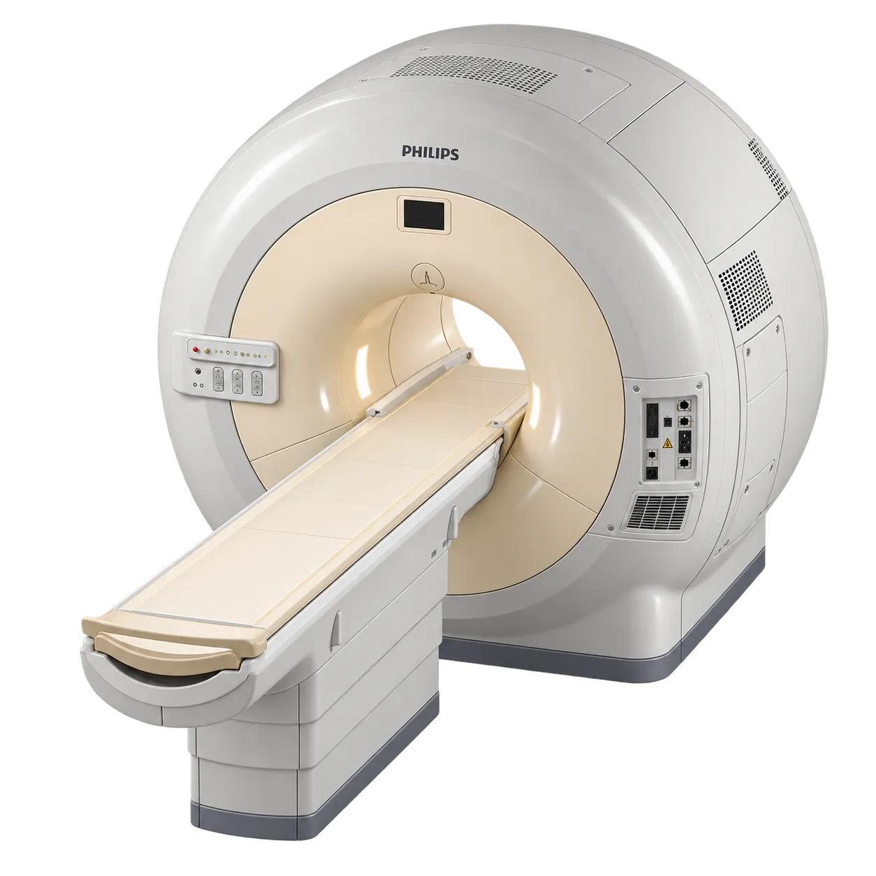
Neurological imaging is supported by high-matrix acquisition and by homogeneity that holds spectrally selective fat suppression across the imaged volume.
Musculoskeletal imaging benefits from matrices to 2,000 by 2,000 and from the gradient performance required for short echo times on the higher amplitude configurations.
Body and abdominal imaging is served by large and offset fields of view, with the body coil supporting large field acquisitions without surface coils where a study permits.
Angiographic work is supported by 4D-TRAK time-resolved acquisition, with gradient performance governing the minimum TE and TR achievable.
Cardiac imaging draws on k-t BLAST acceleration for dynamic studies and on the gradient system fitted to the individual unit.
Oncological staging is supported by whole-body diffusion-weighted imaging with background suppression, acquired across stations on one visit.
Departments running comparable follow-up studies are served by SmartExam with ExamCards, which plans and executes to a stored protocol so examinations match across operators and across visits.