
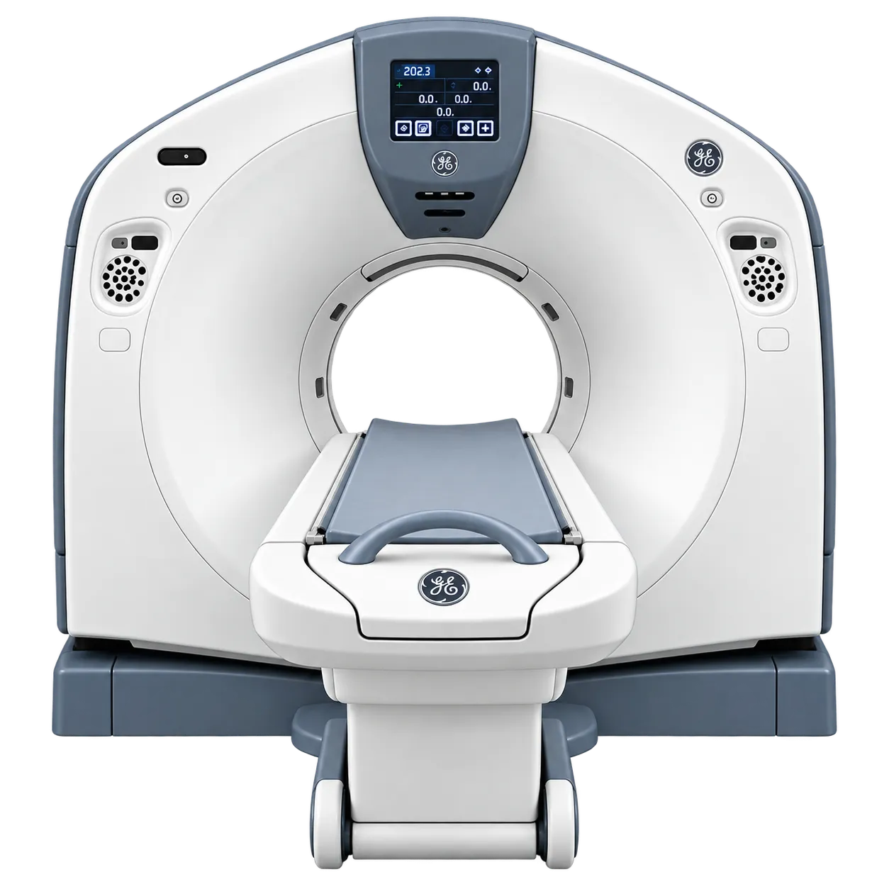
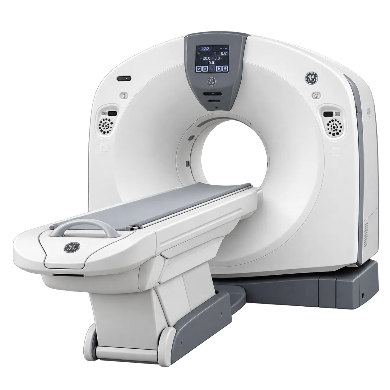
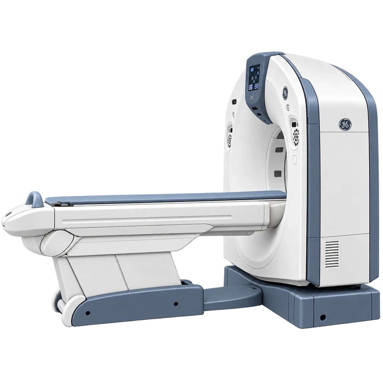
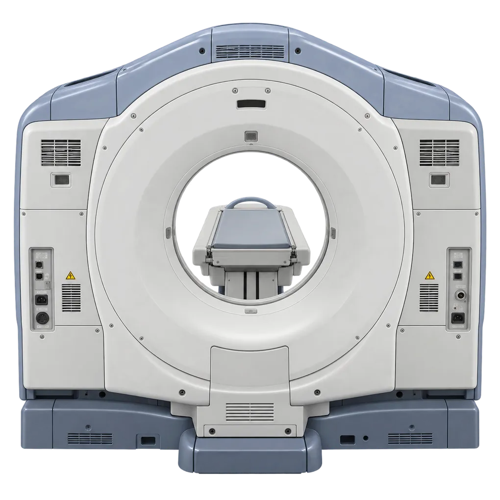
Routine head computed tomography is supported by gantry tilt to 30 degrees, which remains standard practice for angled acquisition in many departments.
Spine imaging uses the same tilt capability alongside sub-millimetre slice thickness for reformatting along the curvature of the spine.
Chest imaging is served by the chest kernel, since a single reconstruction covers both lung and mediastinal review by adjusting window settings.
Abdominal and pelvic work runs across the 50 cm scan field of view with kV selectable to the patient.
Follow-up and surveillance imaging benefits from table positional repeatability within a quarter of a millimetre, so slices can be matched to an earlier study.
High-volume outpatient lists are supported by anode heat dissipation at 820 kHU per minute, which governs how many acquisitions run consecutively.
Emergency presentations are supported by an emergency patient mode for cases where scanning must begin before registration is complete.