
Introduction
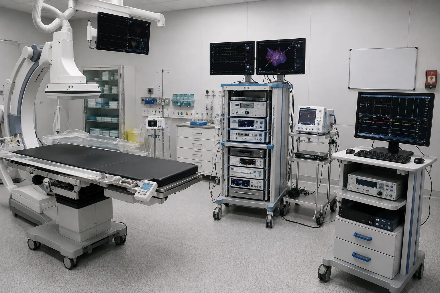
Cardiac electrophysiology deals with the heart's electrical system—the wiring that sets its rhythm—and the abnormal rhythms that arise when that wiring misfires. In an electrophysiology, or EP, study, catheters are placed inside the heart to record its electrical signals, provoke and locate an arrhythmia, and often treat it in the same sitting by ablation. It is a precise, technology-dense discipline in which recording, mapping, ablation and imaging must all work together in one room.
Equipping an EP lab spans the recording system that captures the heart's signals, the electroanatomical mapping that builds a three-dimensional picture of the chamber, the ablation equipment that treats the arrhythmia, the catheter inventory that reaches and works inside the heart, and the imaging and radiation protection around it. This electrophysiology equipment buyer guide sets out what each area needs, how they connect, and the radiation-safety and traceability questions to settle early. It is written for electrophysiologists, cardiac physiologists, EP nurses and procurement across the UK, Europe, the US, the Gulf and South Africa.
What an EP lab needs — the short version
-
An EP recording system and a programmable stimulator to capture and provoke rhythms.
-
Electroanatomical mapping and ablation equipment to locate and treat the arrhythmia.
-
A broad catheter inventory, with imaging and radiation protection around the procedure.
Start with recording and mapping
At the heart of an EP lab is the recording system, which captures the heart's electrical signals in fine detail from catheters placed inside it, alongside a programmable stimulator that paces the heart to provoke and study an arrhythmia. Together they let the team diagnose exactly what rhythm disturbance is present and where it comes from. This diagnostic core is specified first, because everything else in the lab is built around understanding the arrhythmia precisely.
Modern EP work then adds three-dimensional electroanatomical mapping, which combines catheter positions with electrical data to build a colour map of the chamber and the arrhythmia within it. Mapping guides ablation to the right spot with less reliance on X-ray. The mapping platform, iits compatiblecatheters d andthe ablation system are chosen together, bbecausethey form a connected workflow from diagnosis to treatment.
What electrophysiology needs
-
EP recording system. Multichannel capture of intracardiac and surface electrical signals.
-
Programmable stimulator. Controlled pacing to provoke and characterise arrhythmias.
-
Electroanatomical mapping. Three-dimensional mapping of the chamber and the arrhythmia.
-
Ablation system. Radiofrequency or cryoablation to treat the arrhythmia at its source.
-
Catheter inventory. Diagnostic, mapping and ablation catheters in a range of designs.
-
Intracardiac imaging. Intracardiac echo is used to guide access and positioning.
-
Fluoroscopy integration. Imaging support with dose management for catheter guidance.
-
Radiation protection. Shielding, aprons and monitoring for the lab team.
The equipment checklist
|
Equipment |
Role in the lab |
Key selection point |
|
EP recording system |
Signal capture |
Channels; resolution; integration |
|
Programmable stimulator |
Provoking arrhythmia |
Protocols; control |
|
Mapping system |
3D localisation |
Accuracy; catheter compatibility |
|
Ablation generator |
Treatment |
Energy type; catheter range |
|
Catheter inventory |
Access and treatment |
Range; sizing; traceability |
|
Intracardiac echo |
Access guidance |
Image quality; workflow |
|
Radiation protection |
Team safety |
Shielding; monitoring |
Mapping and ablation as one workflow
What defines a modern EP lab is the tight link between mapping and ablation. The three-dimensional map shows the team where the abnormal circuit lives, and the ablation catheter delivers energy — radiofrequency heat or cryoablation cold — precisely to that point to interrupt it. Because the map, the catheters and the ablation generator must be compatible and work as one system, they are planned together rather than bought piecemeal. This connected workflow turns a diagnostic study into treatment in the same procedure.
Imaging and radiation in the EP lab
EP procedures have traditionally relied on fluoroscopy to guide catheters, so the lab operates within the same radiation-safety framework as other X-ray-guided work. Three-dimensional mapping and intracardiac echo now reduce the X-ray needed, but dose management and radiation protection remain essential — pulsed imaging, shielding, aprons and monitoring protect a team that spends long procedures beside the source. Planning imaging and protection alongside the recording and mapping systems keeps the lab both capable and safe.
Compliance and documentation
Electrophysiology equipment must be genuine, current and fully documented, and the lab uses ionising radiation. In the UK, the MHRA regulates medical devices, and NICE publishes clinical guidance. The HSE regulates ionising radiation in the workplace, and medical exposures fall under IR(ME)R, overseen by the CQC. Requirements vary by country and market, so confirm current obligations with the relevant authorities before you buy.
Support keeps an EP lab running.
-
Installation, commissioning and clinical familiarisation for recording, mapping and ablation.
-
Preventive maintenance and checks that keep a connected system dependable.
-
Reliable catheter and consumable supply across diagnostic, mapping and ablation work.
-
Rapid technical response, because a fault interrupts a complex, staffed procedure.
-
Clear documentation and traceability for systems and every catheter used in a case.
More from Business
-
Buyers — source compliant, fully documented equipment.
-
Suppliers — list your equipment and reach buyers worldwide.
-
Advertise — promote your products and services to a clinical audience.
-
Business overview — explore everything MediGear offers healthcare organisations.
Disclaimer
This article is for general information only. It does not constitute clinical, procurement, legal or regulatory advice. Equipment requirements for electrophysiology vary by lab, caseload, facility, country and market, and change over time. The use of ionising radiation is separately governed and requires specialist input. Always confirm current requirements with the relevant authorities, and consult qualified clinical, physiology, biomedical engineering, procurement and regulatory professionals for decisions specific to your organisation. medigear.uk accepts no liability for actions taken based on this content.
"Map it, then treat it." An EP lab equipped for diagnosis and ablation.
We supply medical equipment to healthcare organisations across the UK, Europe, the US, the Gulf and South Africa. Everything is genuine, documented and supported, so your electrophysiology lab diagnoses and treats safely.
Frequently Asked Questions
An EP lab needs a recording system to capture the heart's electrical signals, a programmable stimulator to provoke arrhythmias, and three-dimensional electroanatomical mapping to locate them. It needs an ablation system — radiofrequency or cryoablation — and a broad inventory of diagnostic, mapping and ablation catheters. Intracardiac echo, fluoroscopy integration with dose management, and radiation protection complete the lab.
Electroanatomical mapping combines catheter positions inside the heart with the electrical signals they record to build a three-dimensional, colour-coded picture of the chamber and the arrhythmia within it. This guides ablation precisely to the abnormal circuit and reduces reliance on X-ray. The mapping platform and its compatible catheters are chosen alongside the ablation system as one workflow.
Once mapping locates the abnormal electrical circuit, an ablation catheter delivers energy to interrupt it—radiofrequency uses heat, while cryoablation uses cold. Because the map, catheters and ablation generator must be compatible and work together, they are planned as a connected system. This lets a diagnostic study and its treatment happen in the same procedure.
EP procedures have traditionally used fluoroscopy to guide catheters, so the lab works within a radiation-safety framework. Three-dimensional mapping and intracardiac echo now reduce the X-ray needed, but dose management and protection remain essential because the team spends long procedures beside the source. Pulsed imaging, shielding, aprons and monitoring are planned alongside the recording and mapping systems.
Yes, and it uses ionising radiation. Systems and every catheter used should be genuine, current and traceable. In the UK, the MHRA regulates devices, NICE provides guidance, the HSE oversees workplace ionising radiation, and the CQC oversees medical exposures under IR(ME)R. Requirements vary by country and market, so confirm current obligations with the relevant authorities before purchasing.
Yes. MediGear supplies medical equipment to healthcare organisations across the UK, Europe, the US, the Gulf and South Africa. As a distributor,, we provide genuine, documente,d and supported equipment, from recording and mapping systems to ablation generators, catheter,s and radiation protection. Requirements differ by lab and market, and radiation use is governed, so we work with your team to match equipment to your lab and its regulatory setting.