
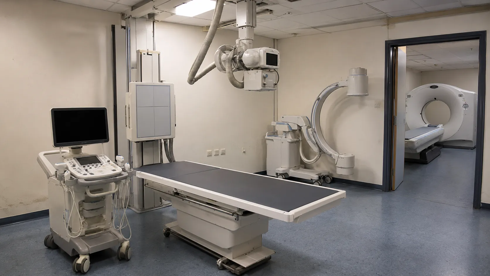
Introduction
Almost every complex diagnosis involves imaging, and imaging depends on the right equipment. Medical imaging spans modalities that use radiation, such as X-ray and CT; those that use magnetic fields or sound waves, such as MRI and ultrasound; and nuclear techniques that show how the body works. Understanding those families helps any service plan, compare options and budget sensibly.
This guide walks through the main types of medical imaging equipment used worldwide, what each does, where it fits, and how to choose — with notes on approval across the UK, the EU, the US, the Gulf (GCC) states, and South Africa.
The short version — imaging equipment in three families
Most imaging equipment falls into one of three families — radiation-based imaging, non-ionising imaging, and functional or molecular imaging.
-
Radiation-based — X-ray, CT, mammography and fluoroscopy, using ionising radiation to see inside the body.
-
Non-ionising — MRI and ultrasound, imaging soft tissue and more without radiation.
-
Functional & molecular — nuclear medicine such as PET and SPECT, showing how the body works.
Types of medical imaging equipment
-
X-ray & digital radiography. Fixed and mobile X-ray systems for bones, the chest and fast triage — the most common imaging.
-
Computed tomography (CT). Cross-sectional X-ray scanners for trauma, stroke and detailed internal imaging in seconds.
-
Magnetic resonance imaging (MRI). Magnetic-field scanners giving the richest soft-tissue detail of the brain, joints and organs.
-
Ultrasound. Sound-wave systems for obstetrics, the abdomen, vascular work and bedside imaging, with no radiation.
-
Mammography. Dedicated low-dose X-ray systems for breast screening and diagnosis.
-
Fluoroscopy & C-arms. Real-time X-ray for theatre, interventional and contrast studies.
-
Nuclear medicine. PET, SPECT and gamma cameras that image function and metabolism, widely used in oncology and cardiology.
-
Bone densitometry (DEXA). Low-dose scanners that measure bone density to assess osteoporosis.
What each type does
|
Category |
Examples |
Used for |
|
X-ray |
Digital radiography, mobile X-ray |
Bones, chest, fast triage |
|
CT |
CT scanners |
Cross-sectional, trauma, stroke |
|
MRI |
MRI scanners |
Soft tissue, brain, joints |
|
Ultrasound |
Diagnostic & Doppler ultrasound |
Obstetrics, abdomen, bedside |
|
Mammography |
Digital mammography |
Breast screening |
|
Fluoroscopy |
C-arms, fluoroscopy units |
Real-time, theatre, interventional |
|
Nuclear medicine |
PET, SPECT, gamma cameras |
Function and metabolism |
|
Densitometry |
DEXA scanners |
Bone density (osteoporosis) |
X-ray vs CT vs MRI vs ultrasound
Imaging is the largest diagnostic family, and the four most common modalities each answer different clinical questions.
|
Modality |
How it works |
Best for |
Consider |
|
X-ray |
Ionising radiation |
Bones, chest, fast triage |
Low cost; radiation dose |
|
CT |
Cross-sectional X-ray |
Trauma, detailed internal imaging |
Higher dose and cost |
|
MRI |
Magnetic fields, no radiation |
Soft tissue, brain, joints |
Costly, slower, contraindications |
|
Ultrasound |
Sound waves, no radiation |
Obstetrics, abdomen, bedside |
Operator-dependent; affordable |
No single modality is best — X-ray and ultrasound are affordable first-line workhorses, while CT and MRI answer more complex questions at higher cost, and most services run several together.
Building imaging capacity?
We'll help you choose the right kit. Tell us the studies, your budget and your market — we'll help you pick imaging equipment that fits your service and is approved for use where you'll use it.
No pressure, no obligation. → Talk to the team
Imaging equipment by setting
-
Large hospitals - the full range from X-ray and ultrasound to CT, MRI and nuclear medicine for complex and emergency care.
-
Imaging & diagnostic centres - X-ray, ultrasound, CT and often MRI, focused on outpatient diagnosis.
-
Clinics & primary care - X-ray and ultrasound, and increasingly handheld ultrasound, close to the patient.
-
Emergency & bedside - portable X-ray and handheld ultrasound for fast answers where the patient is.
-
Screening programmes - mammography and DEXA used at scale for early detection.
Choosing imaging equipment
-
Define the studies and volumes the service must support.
-
Match modalities to the clinical questions and patient mix.
-
Weigh fixed vs portable, and new vs refurbished, against budget.
-
Confirm market approval, the device risk class and radiation safety.
-
Plan shielding, installation, PACS and service support.
Imaging is capital-heavy, and installation, shielding and service contracts add to the purchase price — weigh new against certified refurbished, confirm the device's risk class and radiation requirements, and plan PACS and connectivity before you buy.
Emerging imaging technology
-
AI-assisted imaging. Software flags findings on X-ray, CT and retinal images, speeding reporting and supporting, not replacing, clinicians.
-
Portable & handheld. Pocket and cart ultrasound and lightweight X-ray bring imaging to the bedside and the community.
-
Low-dose detectors. Newer detectors cut radiation dose while improving image quality.
-
Cloud PACS & teleradiology. Images are shared and reported remotely, supporting telemedicine.
-
Hybrid imaging. PET-CT and PET-MRI combine structure and function in a single study.
Approval by region
|
Region |
Regulator |
Requirement |
|
United Kingdom |
UKCA mark (CE accepted to 2028–2030) |
|
|
European Union |
CE marking under the EU MDR |
|
|
United States |
510(k) clearance or PMA approval |
|
|
Gulf (GCC) |
SFDA & GCC bodies |
National registration / GCC conformity |
|
South Africa |
Medical device licensing |
Imaging equipment also follows radiation-safety rules for ionising modalities such as X-ray, CT and fluoroscopy. For clinical and quality guidance, the NICE and World Health Organisation resources are useful references. Our buyer hub sets out how to purchase with confidence.
More from Business
-
Buyers — source imaging equipment, from ultrasound to CT and MRI, for your service.
-
Suppliers — list your imaging and radiology equipment and reach buyers worldwide.
-
Advertise — promote your products and services to a clinical audience.
-
Business overview — explore everything MediGear offers healthcare organisations.
Disclaimer
This article is for general information only and does not constitute clinical, radiological, medical, procurement or regulatory advice. Imaging modalities, requirements and regulations vary by country, clinical setting and need, and change over time. Always confirm that equipment suits the intended clinical use and is approved for your market (for example, UKCA, CE under the EU MDR, FDA, SFDA/GCC or SAHPRA), observe radiation-safety rules for imaging, and consult qualified radiology, medical-physics, biomedical engineering and procurement professionals for decisions specific to your organisation. medigear.uk accepts no liability for actions taken based on this content.
The right imaging for your service.
From X-ray and ultrasound to CT, MRI, mammography and nuclear medicine, we supply imaging equipment to clinics, hospitals and diagnostic centres across the UK, Europe, the US, the Gulf and South Africa — compliant, supported and ready to use.
Frequently Asked Questions
The main types are X-ray and digital radiography, computed tomography (CT), magnetic resonance imaging (MRI), ultrasound, mammography, fluoroscopy and C-arms, nuclear medicine such as PET and SPECT, and bone densitometry (DEXA). X-ray and ultrasound are the most widely used, while CT, MRI and nuclear imaging serve more complex or specific roles.
CT uses X-rays to build detailed cross-sectional images quickly, making it ideal for trauma, stroke and chest or abdominal imaging, but it involves a radiation dose. MRI uses magnetic fields and radio waves, with no ionising radiation, and gives the best soft-tissue detail for the brain, joints and organs, but it is slower, costlier and unsuitable for some patients.
Ultrasound and MRI do not use ionising radiation. Ultrasound uses high-frequency sound waves and is portable and affordable, ideal for obstetrics, the abdomen and bedside use. MRI uses magnetic fields and is the best modality for soft tissue. X-ray, CT, mammography and fluoroscopy all use ionising radiation and follow radiation-safety rules.
Yes. X-ray, ultrasound and even CT and MRI systems are widely available as certified refurbished units and can significantly reduce capital cost. Confirm certified refurbishment, a warranty, installation and service support, and check the device is approved for your market. Scanners need acceptance testing and quality assurance after installation.
Imaging devices must be approved for the market — UKCA or CE under the EU MDR in the UK and EU, FDA clearance or approval in the US, and SFDA or SAHPRA registration in the Gulf and South Africa. Ionising modalities also follow radiation-protection regulations, so plan for shielding, dosimetry, acceptance testing and servicing.
A small clinic or primary-care setting typically starts with X-ray and ultrasound, which together cover a wide range of common questions affordably, and increasingly uses handheld ultrasound. Larger centres add CT and MRI, while mammography and nuclear medicine are usually found in hospitals and dedicated screening services.