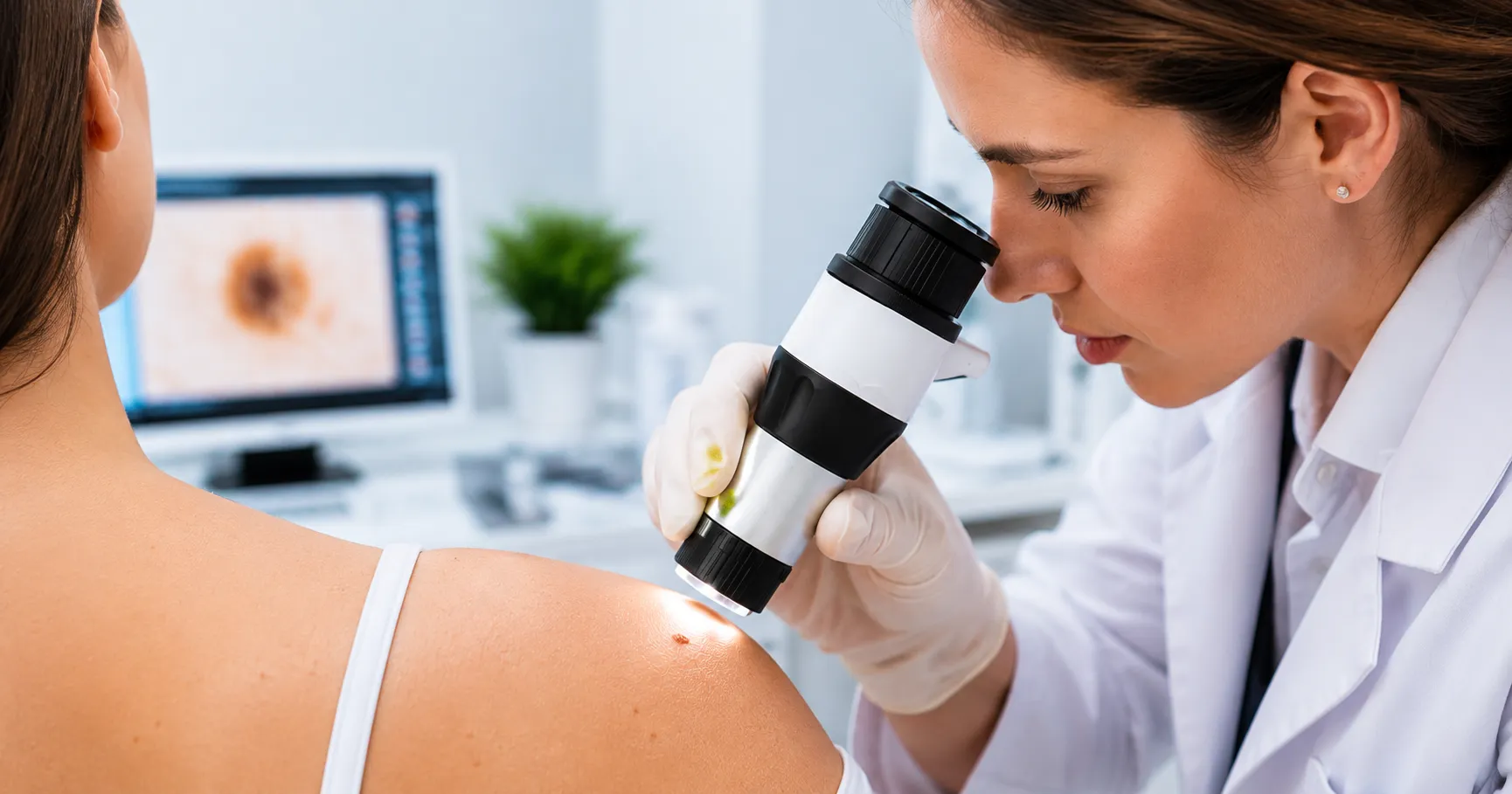
What separates the dermatologist who removes a lesion when not needed from the one who spots the melanoma? The one that looks benign to the eye. In many cases, the dermatoscope. One device. The view the naked eye cannot give. A handheld optical device. Eliminates the air-skin interface. Magnifies. Reveals subsurface structures the naked eye cannot access. Without dermoscopy, a dark spot. With it — a network. A pattern. A structure. The pattern decides. Safe to observe or urgent to excise.
She was forty-three. Pale skin, Irish heritage, history of sunburn in childhood. Came for a mole check. The nurse used the naked eye. Nothing alarming. Routine. The dermatologist used the dermatoscope. Atypical pigment network. Regression. Uneven streaks at the border. All in one view. Melanoma. Early — Clark level two. Excised with clear margins. She did not know. The dermatoscope did.
This guide covers what a dermatoscope is and how dermatologists use it. Honest detail. The kind clinicians, trainees, and procurement teams need. Medigear supplies certified dermatoscopes to dermatology departments, GP surgeries, and skin clinics across the UK. Every point here comes from real clinical demand. From real use. Clinics sourcing certified dermatoscopes can explore the Medigear buyers portal for pricing, availability, and procurement built for dermatology purchasing.
What Is a Dermatoscope
A dermatoscope — also called a dermoscope — is a handheld optical instrument. Used to examine skin lesions. Combines zoom — typically ten times. Illumination. Elimination of surface reflection. Two ways to eliminate this reflection. Each has its role. Contact dermoscopy uses fluid. Gel, oil, or alcohol. Applied between the lens and the skin. Polarised dermoscopy uses cross-polarised light. No contact. No fluid. Each reveals what the other can miss. Polarised — clear vascular structures. Contact fluid — scale and surface detail.
Dermoscopic Structures
The structures the dermatoscope reveals are the language of dermoscopy. Pigment network — honeycomb. Benign naevi — regular, fading. Melanoma — uneven mesh, abrupt endings. Dots and globules — melanocyte clusters. Regular — benign. Uneven — suspicious. Streaks — radial projections at the border. Asymmetric ones raise concern. Regression structures — white scar areas. Blue-grey peppering. Both associated with melanoma. Vascular structures — vessel patterns. Regular dots — benign. Polymorphous — concerning. It gives the vocabulary. The clinician makes the call. Dermatoscope manufacturers wanting to list polarised, contact, and video systems where dermatology clinics and GP surgeries are searching can reach buyers through the Medigear advertising platform.
Handheld Devices
Handheld devices are the most common type. LED illumination. Polarised, non-polarised, or switchable. Battery-powered. Pocket-sized. Used close to the skin. Held to the lesion. Examined on screen or eyepiece. Simple. No setup. The dermatoscope, always in the pocket, gets used at every consultation. The one requiring setup — referrals only. Reach out to our team for guidance on matching dermatoscope type to your clinical setting and patient volume.
Video Dermatoscopes
Video devices — dermoscopy cameras. Connect to a monitor, tablet, or computer. Image on screen. Captured. Stored. Compared at six or twelve months. Total-body photography provides a baseline for every lesion. Regular monitoring detects change. Early. Before it crosses into something urgent. Mapped today. A comparison for every future review. Change becomes visible.
Non-Pigmented Uses
The device is also used for non-pigmented conditions. Hair and scalp — reveals shaft, follicle openings, scaling, vessel patterns. Many conditions are named without biopsy. Nail disorders — nail fold capillaries show systemic vascular conditions. Inflammatory conditions — psoriasis, lichen planus, sarcoidosis. Each has its own features under the lens. Infestations — the scabies burrow and the mite itself become visible. Vascular lesions — haemangiomas, pyogenic granulomas, angiokeratomas. Each shows a typical pattern. Our guide to diabetes and medical devices covers how monitoring tools transform the management of chronic conditions — the same principle applies when dermoscopy transforms the management of skin lesions that require monitoring rather than immediate excision.
Two-Step Method
The two-step method is the starting point. Used with a dermatoscope in skin lesion assessment. Step one. Is this melanocyte-based? No — check for non-melanocyte patterns. Yes — go to step two. Benign, suspicious, or malignant? Use the ABCD rule, seven-point checklist, or Menzies method. Different clinicians, different methods. All need knowledge of the structures and experience reading images. Our guide to myasthenia gravis covers how diagnostic precision depends on using the right tool in the right hands — the same principle applies when the dermatoscope in the hands of a trained clinician changes the outcome of a lesion that would otherwise be dismissed or removed when not needed.
GP Triage
Can your GP surgery offer dermoscopy triage before referring to secondary care? A trained GP can tell the urgent two-week-wait from the six-month review. The dermatoscope makes that call possible. Without one — refer everything odd. Flood the pathway. Lesions dismissed in ten seconds. Suppliers of handheld, polarised, and video systems can register through the Medigear supplier portal to connect with GP surgeries, skin clinics, and dermatology departments investing in dermoscopic capability.
Choosing a Device
What should a GP look for when choosing a dermatoscope for primary care? First — polarised or contact, or switchable? Switchable gives the most versatility. Second — eyepiece or screen? Screen-based devices allow the patient to see the image, which is useful for shared decision-making. Third — standalone or smartphone-connected? Adapters cost less. Good for low volume. Fourth — video capture? For lesions monitored over time, stored images are essential. The one that fits the workflow gets used. The one that does not gets put away.
Protocol
Does your dermatology unit have a protocol? Same lighting every time. Same zoom level. Same record for every lesion reviewed. Consistent protocol — meaningful comparison. Done each time differently — images that cannot be compared. Review that starts from scratch.
Seven-Point Checklist
Can your team identify features that make a pigmented lesion suspicious? And know when the dermatoscope shows enough to refer? Without waiting for more? Seven-point checklist: atypical network, blue-white veil, irregular streaks, irregular pigment, irregular dots, regression, atypical vessels. Three minor criteria — or any major one — warrant referral. Better information for the clinician. The decision stays theirs.
Many Moles
How does your clinic manage the patient with many moles and one that has changed? Total body photography gives the baseline. The video device gives the comparison. The lesion that has grown, changed colour, or developed new structures between visits — found before it becomes the melanoma it is about to be.
Mole Map
Can your team check a mole map in order — working through zones of the body so no area is missed? A structured check — scalp, face, ears, neck, chest, back, arms, hands, legs, feet. Same order each time. No awkward site skipped. The back and scalp — patients cannot self-examine there. Also exactly where melanomas are found late. The dermatoscope reaches those sites.
Rule of Two
What does your clinic do when a lesion has features hard to classify — not clearly benign and not clearly malignant? The rule of two is a useful guide. In doubt after dermoscopy? Refer it. The cost of a not needed referral is low. The cost of a missed melanoma is not.
Sequential Imaging
Does your team use regular imaging for any patient with more than fifty moles, a history of melanoma, or a first-degree relative who has had melanoma? These patients have a higher risk. A single check is not enough. Regular monitoring — baseline images compared to follow-up at six or twelve months — finds the lesion that has changed while it is still early. Change over time is often more telling than appearance on a single day.
Image Storage
How does your clinic store and review dermoscopy images across visits? Images stored in the patient record allow any clinician to review baseline and current images side by side. Images taken and not stored are lost. No comparison. The lesion reviewed without the previous image starts from scratch. The change goes unseen. The one that mattered.
Training
Does your team receive structured dermoscopy training alongside the dermatoscope — or just the device? Without training, the device makes a clinician more confident. In all the wrong answers. A two-day course and supervised practice. That converts it into a diagnostic upgrade. Buy without training. Miss the melanoma the same way the naked eye did. Different zoom. Same result. Companies seeking long-term collaboration on supply, training, and skin lesion monitoring can explore the Medigear partnership programme for ongoing opportunities.
Why Choose Medigear
Medigear supplies certified handheld, polarised, and video systems to dermatology departments, skin clinics, and GP surgeries across the UK. Whether equipping a new skin clinic, adding dermoscopy to an existing service, or building a GP-led triage pathway — our team matches the right device to your setting and patient population. Reach out to our team for guidance built around the structures that tell the clinician what the naked eye cannot — and the instrument that makes them visible.
Conclusion
What separates the dermatologist who spots the melanoma from the one who dismisses it? The dermatoscope. She was forty-three. Came for a mole check. Nurse — nothing alarming. Dermatologist used the dermatoscope. Atypical network. Regression. Uneven streaks. Melanoma. Clark level two. Excised. Survived. She did not know. The device did. Polarised or contact. Handheld or video. Switchable or dedicated. Pigment network. Dots. Regression. Streaks. Vessels. Two-step method. ABCD rule. Seven-point checklist. Menzies. GP triage to separate the urgent from the watchable. Protocol for every examination. Images stored and compared. Training alongside the device — not instead of it. Medigear stands alongside dermatology teams with certified dermatoscopes matched to the setting and the patient. Speak to our team today — because the structure that tells the clinician what the naked eye cannot see must be visible through the right device, in the right hands, at the right time.
⚠️ This post is for general information only. We do not sell medications or provide prescriptions — Medigear.uk is a medical equipment supplier only.