
Introduction
Cardiology generates a remarkable amount of data about the heart — the traces of its electrical activity, the moving pictures of its chambers, the scans of its structure. Reading it all well and drawing the right conclusions is demanding expert work, and it is exactly the kind of pattern-rich task where artificial intelligence is transforming the equipment cardiology relies on.
This article explains how AI is transforming cardiology diagnostic equipment in hospitals — interpreting the heart's electrical traces, reading its moving images, and measuring its structure and function. It is written for cardiology, imaging and procurement teams across the UK, Europe, the US, the Gulf and South Africa who want to understand where AI genuinely helps in diagnosing the heart. (For continuous cardiac monitoring specifically, see our separate guide.)
The short version
-
Cardiology produces rich data: electrical traces, moving images and scans.
-
AI helps interpret ECGs, read heart imaging and measure structure and function.
-
It brings speed and consistency to demanding, expert reading.
-
It supports the cardiologist's judgement rather than replacing it.
Reading the electrical trace
The electrocardiogram, or ECG, is one of medicine's most common tests, recording the heart's electrical activity, and interpreting it well is a real skill. AI trained on enormous numbers of ECGs can help read them—recognising patterns of various conditions and flagging abnormal traces for a clinician's attention, quickly and consistently.
What is striking is how much AI can sometimes draw from an ECG. Beyond the obvious rhythm abnormalities, models have shown an ability to detect subtler signs that a human reader might not, hinting at conditions not traditionally read from a trace. This must be handled with care and proper evidence, but it suggests the humble ECG, read by AI, may hold more information than long assumed — a genuinely intriguing development.
Making sense of the moving heart
Much of cardiology is imaging, and the echocardiogram — an ultrasound of the beating heart — is central to it. Reading these moving images, and measuring the heart's chambers and function from them, is skilled and somewhat variable work. AI can help by automating the measurements, identifying the right views, and quantifying how well the heart is pumping, consistently and quickly.
This consistency matters because so many cardiac decisions rest on these measurements, and manual measurements vary between operators. Automated, reproducible measurement gives a steadier basis for decisions and frees the cardiologist to focus on interpretation and judgement rather than the painstaking work of measuring. It makes a demanding imaging task faster and more reliable.
Where AI helps in cardiology diagnosis
|
Task |
What AI adds |
Why it matters |
|
ECG reading |
Recognises trace patterns |
Fast, consistent interpretation |
|
Echocardiography |
Automates measurement |
Reproducible cardiac figures |
|
Cardiac imaging |
Helps read CT and MRI |
Structure assessed clearly |
|
Risk |
Draws data together |
Better-informed decisions |
Seeing the heart's structure
Beyond ultrasound, CT and MRI image the heart, showing its structure and function in great detail, and AI is proving its worth here too. It can help read these complex scans — measuring structures, quantifying features, and highlighting findings — supporting the cardiologist in drawing accurate conclusions from rich, intricate images.
This matters because cardiac imaging is technically demanding, and consistent measurement across the range of scans a hospital performs is hard to achieve by hand. AI that brings speed and consistency to reading cardiac CT and MRI helps the imaging deliver more, and more reliably, which feeds directly into better-founded decisions about a patient's heart.
Bringing the cardiac picture together
Cardiology decisions often draw on many strands — the trace, the images, the measurements, the history — and one of AI's quieter roles is helping bring them together and weigh a patient's risk. By assembling the relevant information and helping estimate the likelihood of problems, it can support the cardiologist in deciding how closely to watch or how actively to treat.
As with any prediction, this is guidance rather than certainty, and a clinician must weigh it against the whole patient. But used well, a clearer view of risk, drawn from all the available cardiac data, can help focus attention and treatment where they will do the most good — which in heart disease, where so much turns on timely action, is genuinely valuable.
Getting cardiology AI right
For a cardiology service adopting AI, a few principles keep it safe and useful:
-
It was validated on the traces, scans and patients you actually see.
-
It performs well across the range of hearts and conditions you assess.
-
It fits your ECG, echo and imaging equipment and workflow.
-
Its approved intended use matches how you intend to use it.
-
The cardiologist interprets and decides, with the AI in support.
Held to cardiac standards
Because cardiology decisions carry serious consequences, the equipment is held to demanding standards, and the AI within it is no exception. When software serves a medical purpose, it becomes a regulated medical device; the FDA oversees cardiovascular devices, and for the AI element, the FDA reviews software based on its intended use, while in Great Britain the MHRA sets out how AI as a medical device is classified. Validation, performance on your patients, and ongoing monitoring remain the key checks.
Faster reporting, shorter waits
A practical benefit that is easy to overlook is speed of reporting. Cardiology tests are performed in large numbers, and reading and reporting can become a bottleneck, leaving patients waiting for results that are ready but not yet interpreted. By helping with reading and prioritising urgent cases, AI can ease that bottleneck, so results reach patients and their doctors sooner.
Shorter waits are not merely a convenience; in heart disease they can matter clinically, because a delayed result can mean a delayed decision about a serious condition. A cardiology service that can turn around tests faster, without cutting corners on quality, serves its patients better, and this quiet gain in throughput is among the more tangible benefits AI brings to a busy department.
The equipment beneath the intelligence
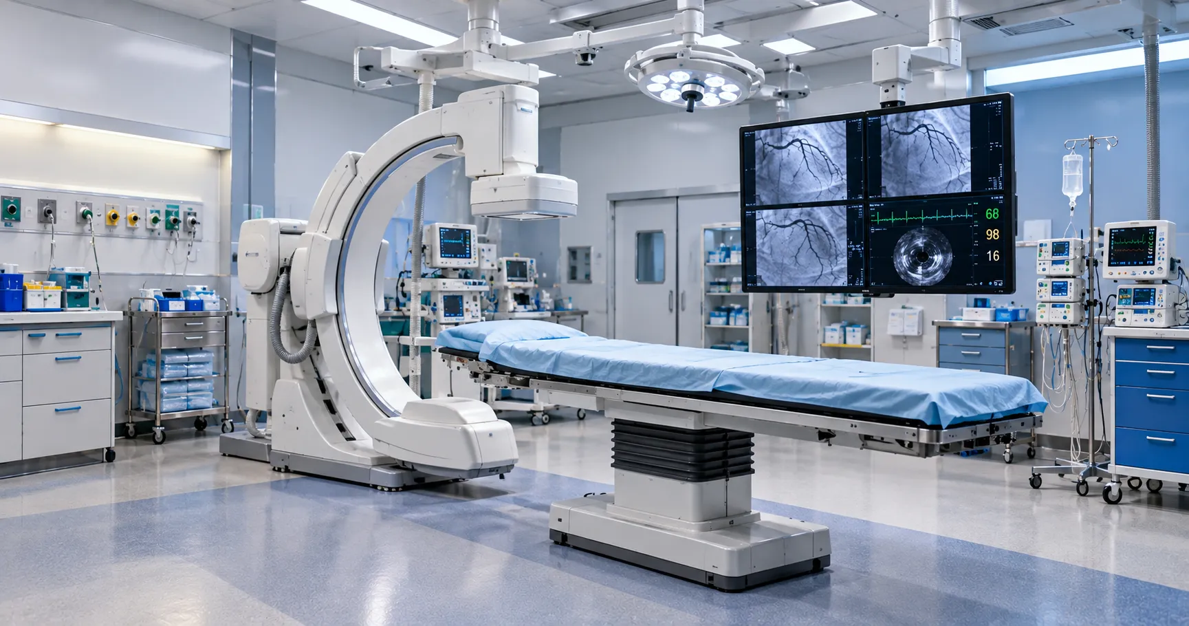
This is why a hospital weighing cardiology AI should look at the whole picture rather than the software alone. The most capable interpretation in the world adds little to a poor image. In contrast, even modest assistance can shine on a clear one, so the equipment and the intelligence are best judged, and best chosen, together as a single, coherent system that reads the heart clearly and well.
It is worth stressing that all of this rests on the quality of the underlying cardiac equipment. An ECG machine that records a poor trace, or an ultrasound that produces an unclear image, gives AI poor material to work with, and no amount of clever interpretation rescues a bad recording. The intelligence is a layer on top of sound diagnostics, never a substitute for them.
So the sensible view is that good cardiology AI and good cardiology equipment go together. Investing in reliable, well-maintained machines that produce clear, accurate recordings is what lets any intelligence layered on top deliver its full value. The heart is read best when capable equipment and capable software work together, each doing what it does well.
A clearer read on the heart
AI is transforming cardiology diagnostic equipment by bringing speed and consistency to the demanding work of reading the heart — interpreting its electrical traces, measuring its moving images, and making sense of its structure on complex scans. It does not replace the cardiologist but gives their expert eye a tireless, consistent partner. Held to proper standards, it helps cardiology see the heart more clearly and decide more surely, which is what good heart care depends on.
More from Business
-
Buyers — source approved, well-documented equipment.
-
Suppliers — list your equipment and reach buyers worldwide.
-
Advertise — promote your products and services to a clinical audience.
-
Business overview — explore everything MediGear offers healthcare organisations.
Disclaimer
This article is general information, not clinical or procurement advice. Cardiology AI should be used within its approved intended use and under qualified clinical supervision; confirm current requirements with the relevant authority. medigear.uk accepts no liability for actions taken based on this content.
Cardiology equipment you can build on
Genuine, approved, well-documented cardiology and imaging equipment — the dependable foundation good cardiac AI reads from, wherever you care for the heart.
Frequently Asked Questions
Cardiology produces rich data — electrical traces, moving images and scans — and AI brings speed and consistency to reading it. It helps interpret ECGs, automate echocardiography measurements, read cardiac CT and MRI, and integrate data to assess risk. It supports the cardiologist's judgement rather than replacing it. (Continuous cardiac monitoring is covered in a separate guide.)
Yes. Trained on enormous numbers of ECGs, AI can recognise the patterns of various conditions and flag abnormal traces for a clinician quickly and consistently. Strikingly, models have shown an ability to detect subtler signs a human reader might not, suggesting the ECG may hold more information than long assumed — though this must be handled with care and proper evidence.
Reading moving ultrasound images of the beating heart and measuring its chambers and function is skilled, somewhat variable work. AI can automate the measurements, identify the right views and quantify how well the heart pumps, consistently and quickly. Reproducible measurements provide a steadier basis for decisions and free the cardiologist for interpretation.
Yes. CT and MRI image the heart in great detail, and AI can help read these complex scans—measuring structures, quantifying features, and highlighting findings—supporting the cardiologist in drawing accurate conclusions. It brings speed and consistency to technically demanding imaging that is hard to measure uniformly by hand.
Cardiac monitoring AI watches continuous signals in real time — catching fleeting arrhythmias and warning of deterioration, covered in a separate guide. This piece is about cardiology diagnostic equipment: interpreting ECGs, reading echo and cardiac imaging, and measuring the heart's structure and function to reach a diagnosis, rather than monitoring a patient.
Yes. When such software serves a medical purpose, it is a regulated medical device; cardiovascular devices are overseen as medical devices, and the AI element is reviewed based on its intended use. Validation of the traces, scans, and patients you see; performance across the conditions you assess; and ongoing monitoring remain the key checks.