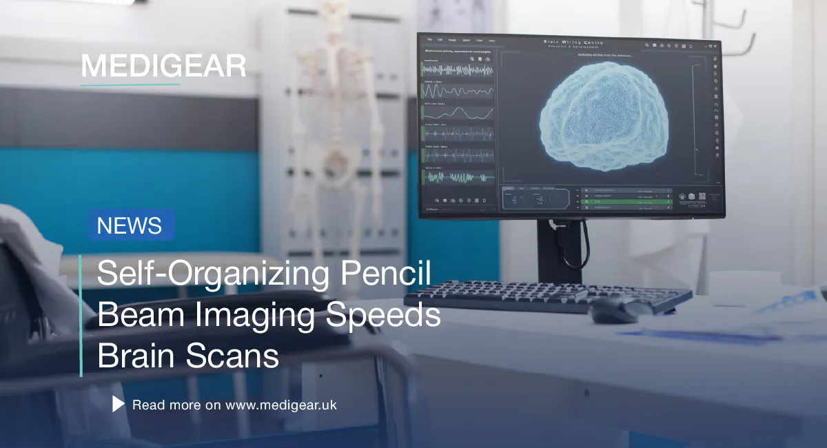
Self-Organizing Pencil Beam Imaging Speeds Brain Scans
A new self-organizing pencil beam imaging method from MIT could speed up drug tracking across the blood-brain barrier. Laser light that should scatter inside an optical fiber snaps into a narrow "pencil beam."
The work, in Nature Methods, used the self-organizing pencil beam imaging to make 3D images of the blood-brain barrier roughly 25 times quicker than today's gold-standard approach. Quality held up. The team can also watch single cells absorb drugs in real time. That helps evaluate whether Alzheimer's and ALS treatments reach their brain targets.
A surprise inside an optical fiber
The finding came from an odd observation. The MIT team had built a precise fiber shaper to steer laser light through a multimode optical fiber. These fibers can carry high power.
Lead author Honghao Cao, an EECS graduate student, raised the power to test the fiber's limits. Wisdom said higher power means more scatter. Instead, near the damage threshold, the light snapped into one sharp beam.
Sixian You, assistant professor in MIT's EECS Department and senior author, said: "The common belief in the field is that if you crank up the power in this type of laser, the light will inevitably become chaotic. But we proved that this is not the case."
You said the method skips most of the optical engineering normally needed at high power.
How self-organizing pencil beam imaging works
The effect needs two conditions. The beam must enter the fiber at exact zero-degree alignment. The power must climb until the light couples with the fiber's glass.
"At this critical power, the nonlinearity can counter the intrinsic disorder, creating a balance that transforms the input beam into a self-organized pencil beam," Cao said.
These conditions are rarely tried together. Labs avoid the highest power to protect the fiber. Tight alignment is not standard either. Pair them and the fiber yields a stable, sharp beam, on a standard optical bench.
Sharper, clearer images
The self-organizing pencil beam imaging is both stable and detailed. Many beams produce "sidelobes" — blurred halos that cut image clarity. The pencil beam stays clean and tightly focused.
The team then aimed the technique at the human blood-brain barrier — a dense cell layer that guards the brain but blocks many drugs.
25x faster blood-brain barrier scans
Drug developers track compounds through blood vessels and into brain tissue. Old optical methods capture one 2D slice at a time, then stitch scans into a 3D image — slow work.
The self-organizing pencil beam imaging method made rapid, high-precision 3D images. The team also tracked how cells absorb proteins in real time. Cellular-level 3D images came out roughly 25 times faster, with improved quality. The new self-organizing pencil beam needs no fluorescent labels — an old barrier in drug-tracking work.
Roger Kamm, Cecil and Ida Green Distinguished Professor of Biological and Mechanical Engineering at MIT, said: "The pharmaceutical industry is especially interested in using human-based models to screen for drugs that effectively cross the barrier, as animal models often fail to predict what happens in humans."
Sarah Spitz, a postdoctoral co-author, said the method extends beyond the barrier. It enables time-resolved tracking of diverse compounds across engineered tissue models.
Resolution and depth, together
Microscopy normally trades resolution for depth of focus. The deeper you image, the lower the resolution.
You said: "Usually, you have a tradeoff between image resolution and depth of focus -- you can only probe so far at a time."
The self-organizing pencil beam imaging beats that trade. It matters most for layered tissue models like the blood-brain barrier.
Funders and next steps
Other authors: EECS grad students Li-Yu Yu and Kunzan Liu. Postdocs Francesca Michela Pramotton and Federico Presutti contributed. Zhengyu Zhang PhD '24 also joined, alongside Subhash Kulkarni of Harvard and Beth Israel Deaconess Medical Center.
Funders: MIT startup funds, the National Science Foundation, the Silicon Valley Community Foundation, the Diacomp Foundation, the Harvard Digestive Disease Core, a MathWorks Fellowship, and the Claude E. Shannon Award.
Next, the team will dig into the physics, extend the method to neurons, and push toward clinical use.
The self-organizing pencil beam imaging could reshape how clinicians screen drugs and probe brain tissue. Coverage on Medigear.uk shows why hospital teams must follow how these findings shape neurology.
Source: Originating coverage based on Massachusetts Institute of Technology press materials by Adam Zewe on the You/Cao et al. paper in Nature Methods. Sixian You, assistant professor in the MIT Department of Electrical Engineering and Computer Science, senior author. Honghao Cao, EECS graduate student, lead author. Roger Kamm, Cecil and Ida Green Distinguished Professor of Biological and Mechanical Engineering, MIT. Sarah Spitz, postdoctoral researcher, MIT. Co-authors include Subhash Kulkarni of Harvard University and Beth Israel Deaconess Medical Center.