What is the chest tightness that appears on the hill and disappears at the bottom? The pressure behind the sternum that arrives with effort and retreats with rest? The symptom the patient dismisses as indigestion, the clinician investigates as cardiac, and the monitor confirms as ischaemia? Angina. The heart muscle demands more oxygen than the narrowed artery can supply. Not a heart attack. Not permanent damage. A warning — vessel narrowed, muscle at risk, next demand may cross into a heart attack.
She was sixty-one. Retired nurse. Chest tightness when climbing stairs. Stopped at the top. Tightness went. Thought nothing of it. Three months later — same on an incline. Then flat ground. Then, making the bed. Came to the GP with indigestion that came and went. Resting ECG — normal. Reassured nobody. Angina is dynamic. Heart at rest in the GP surgery may look normal on the trace. The same heart on the stairs produces ST depression that the resting trace never sees. Referred for a stress test. Exercise ECG showed ST depression at low workload. Angiography — tight stenosis in the LAD. Stented. Resolved. The investigation found what the resting ECG had missed.
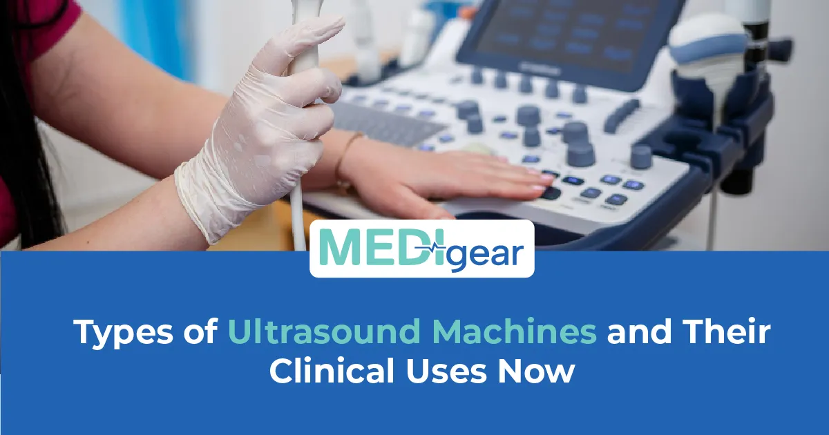
This guide covers what angina is and how cardiac monitors help detect it with the honest detail that GPs, cardiologists, and procurement teams need. Medigear supplies certified cardiac monitors to hospitals, GP surgeries, and specialist clinics across the UK, and every point here comes from real clinical demand. Clinics sourcing certified cardiac monitors can explore the Medigear buyers portal for pricing, availability, and procurement built for cardiac diagnostic purchasing.
What Is Angina
Angina is chest pain or discomfort caused by reduced blood flow to the heart muscle. Coronary arteries supply oxygenated blood to the heart. Fatty plaque narrows an artery. Enough at rest — not enough when demand rises with exercise, stress, or cold. Demand exceeds supply. Ischaemia follows. Symptoms follow ischaemia. Stable angina is predictable. Same trigger. Same duration. Same relief with rest or nitrate. Unstable angina occurs at rest or with minimal effort. The plaque has changed. The vessel is partially blocked. The risk of a heart attack has risen sharply. An acute coronary syndrome. Not stable angina that worsened. A different emergency. Hospitals and cardiology units sourcing certified cardiac monitoring equipment can explore the Medigear buyers portal, a procurement platform built for cardiac diagnostics.
Symptoms
Chest tightness or pressure. Not sharp or stabbing. Radiation to the left arm, jaw, neck, or back. Breathlessness. Nausea. Sweating. Brought on by effort, emotion, cold, or a heavy meal. Relieved by rest within minutes. Relieved by nitrate under the tongue within two to five minutes. Band across the chest. Makes them stop. Passes when they sit down. Classic angina. Stabbing pain with deep breathing — something else. Recognise the character and trigger. The monitor provides the objective evidence. Cardiac monitor manufacturers wanting to list ECG machines, Holter systems, and stress testing equipment where hospitals and GP surgeries are searching can reach buyers through the Medigear advertising platform.
Resting ECG
The resting twelve-lead ECG is the starting point. Often normal in stable disease. May show evidence of a previous heart attack — Q waves, T wave changes, bundle branch block. Or left ventricular hypertrophy. Provides the baseline. A normal resting ECG does not exclude the condition. Starts the investigation, the dynamic tests continue. Reach out to our team for guidance on matching cardiac monitors to your GP surgery or outpatient cardiology setting.
Exercise ECG
The exercise ECG — stress test — is the classic investigation for stable chest pain. Patient walks on a treadmill or cycles. Work rate increases. A continuous ECG is recorded throughout. Clinician watches for ST changes. ST depression of one millimetre or more with exercise and resolving at rest — hallmark of ischaemia. Symptoms at low workload are a poor prognostic sign. Normal capacity with no ST changes, and no symptoms — angina is less likely. The exercise ECG connects the symptom to the objective finding that the resting trace could not see.
Holter Monitor
Holter monitoring records continuously for twenty-four to forty-eight hours while the patient goes about their daily life. Patient presses the event marker when symptoms occur. The clinician reviews the trace at that moment. ST depression at the moment of tightness — ischaemia confirmed. No change — may point to a non-cardiac cause. Holter also finds arrhythmias the patient blames on angina — AF, SVT, ectopics. Our guide to Creutzfeldt-Jakob disease covers the continuous biomedical monitoring standards used in neurological conditions — the same commitment to uninterrupted cardiac monitoring applies when a Holter must capture the ST change the patient pressed the button to report.
CTCA
CT coronary angiography — CTCA — uses contrast and a CT scanner to image the coronary arteries without surgery. Shows the vessel anatomy. The stenosis. The plaque. Whether the narrowing is significant. First-line for stable chest pain in many UK cardiology pathways. Answers the anatomical question — is there a disease? — before asking how the heart responds to demand. Negative CTCA with typical symptoms — excludes significant coronary disease in most cases. Our guide to essential eye screening devices covers the principles of early imaging-based diagnosis in preventing irreversible damage — the same principle applies when CTCA catches the coronary stenosis before the demand that crosses into a heart attack.
Cardiac Perfusion Scan
Cardiac perfusion scan uses a radiotracer to show blood flow through the heart at rest and during stress. Areas with less blood during stress appear as defects on the scan. Functional test. Shows whether the stenosis causes ischaemia — not just that it exists. Used when the anatomy is known but its significance is uncertain. Or when the stress ECG is inconclusive.
Cardiac MRI
MRI provides detailed structural and functional imaging. Wall motion in stress. Perfusion defects. Old heart attack scarring. Anatomy, function, and tissue detail — all in one scan. Used where other tests have not given a clear answer.
Same-Day ECG
Does your GP surgery offer a same-day ECG and referral for stress testing — not waiting weeks for the cardiology outpatient? Exertional tightness with normal resting ECG — the stress test most likely changes this patient's pathway. ECG in the room where the symptom is described initiates the pathway immediately. Booked for next week — seven days lost. Suppliers of ECG machines, Holter monitors, and exercise testing systems can register through the Medigear supplier portal to connect with GP surgeries and cardiology units building their angina detection capability.
Rapid Access Clinic
Does your cardiology team offer a rapid access chest pain clinic within two weeks of GP referral for every patient with suspected stable angina? Three months waiting — the patient may present as an emergency before the appointment arrives. Rapid access. Resting ECG. Stress test or CTCA within two weeks. The fast pathway catches the disease before it catches the patient. Companies seeking long-term collaboration on cardiac monitoring supply, servicing, and cardiology diagnostic packages can explore the Medigear partnership programme for ongoing opportunities beyond a single order.
History First
Can your team separate angina from musculoskeletal chest pain in the history alone? Pain worse with movement, reproduced by pressing on the chest, sharp, not tight, present at rest and with effort, no link to exercise load — less likely to be angina. The tight, exertional, predictable one relieved by rest or nitrate is classic. History first. ECG confirms. Stress test reveals.
Remote Monitoring
Does your cardiology team offer remote monitoring — event recorders, implantable loop recorders, or wearable patches for patients whose symptoms are infrequent and hard to capture? The patient whose symptoms occur once a week is not well served by a 24-hour Holter monitor. The two-week patch or implantable monitor gives the recording window the symptom needs to fall into.
Pattern Change
What does your team do for the patient with known stable disease who reports a change in symptoms at lower thresholds, more frequent, lasting longer? Same-day review. ECG. Troponin. Review medication. Possible same-day referral. The change in pattern warns that the next episode may not resolve.
Patient Education
Does your team educate every newly diagnosed patient about the difference between stable and unstable symptoms — and what to do when the pattern changes? Stable angina — same trigger, same duration, same relief — managed. Angina at rest, more than fifteen minutes, not relieved by two nitrate doses — call an ambulance. A patient who knows the difference acts in time. The one who waits for it to pass may not have time.
Waiting for Revascularisation
How does your team support patients waiting for stenting or bypass? Medical therapy — beta blockers, nitrates, calcium channel blockers, antiplatelet drugs. Lifestyle. Symptom diary. Escalation plan if things worsen. The wait for stenting or bypass is not passive. Managed — right drugs, right monitoring, right instructions for when to call an ambulance.
Why Choose Medigear
Medigear supplies certified resting ECG machines, exercise ECG systems, Holter monitors, and cardiac diagnostic accessories to hospitals, GP surgeries, and cardiology units across the UK. Whether you are equipping a rapid access chest pain clinic, upgrading Holter capability, or building a complete cardiac diagnostic pathway, our team matches the right cardiac monitors to your patients and your practice. Reach out to our team for guidance built around the heart that gives warning — and the monitors that hear it.
⚠️ This post is for general information only. We do not sell medications or provide prescriptions — Medigear.uk is a medical equipment supplier only.