What does an ophthalmologist see that a GP cannot? The answer is not knowledge. It is the equipment. Slit lamp — cornea at forty times. Tonometer — eye pressure. Fundus camera — retina captured. OCT — macula in microns. Visual field — maps what the patient sees and misses. Without the right eye screening devices, the clinician is limited, like anyone else. With them, glaucoma is found before the patient notices. Retinopathy caught before the bleed. Macular changes spotted before central vision goes. Eye screening devices are the exam. Not just a help.
She set up a private ophthalmology clinic. Two consulting rooms. One procedure room. Long equipment list. Short budget. Tough choices ahead. Picked the eye screening devices covering the most ground in the least time. Slit lamp. Tonometer. Autorefractor. Fundus camera. OCT. Visual field analyser. Phoropter. Seven devices. Ninety per cent of clinical need on day one. Pachymeter, specular microscope, topographer — later, as the case mix grew. Clinic opened on time. Patients came from week one. The eye screening devices paid for themselves before the first quarter ended.
This guide covers the essential eye screening devices every ophthalmologist needs with the honest detail that clinic owners, ophthalmic procurement teams, and clinical leads need. Medigear supplies certified eye screening devices to ophthalmology practices across the UK, and every point here comes from real clinical demand. Clinics sourcing certified eye screening devices can explore the Medigear buyers portal for pricing, availability, and procurement built for ophthalmic purchasing.
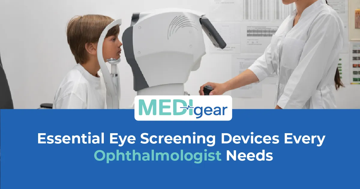
Slit Lamp
The slit lamp is the starting point. Every ophthalmic examination begins here. Binocular microscope. Focused beam of light. Cornea. Chamber. Iris. Lens. Vitreous. Extra lenses — retina and disc. Six to forty times. LED. Beam adjustable — width, height, angle. Shows what the naked eye misses. Quality optics matter here. Used on every single patient who walks through the door.
Tonometer
The tonometer measures intraocular pressure — the number that screens for glaucoma. Goldmann applanation — gold standard. Mounted on the slit lamp. Measures the force to flatten the cornea. Air puff — quick, less accurate. Rebound — handheld, no drops, suits children. Miss the pressure — miss the glaucoma. Catch it — treat before the optic nerve damage sticks. Reach out to our team for guidance on matching eye screening devices to your clinical case mix and patient volume.
Autorefractor
The autorefractor and keratometer measure refractive error and corneal curvature. Objective. Fast. Patient sits. Device measures. Starting point in seconds. Combined units save space and cost. Three seconds versus three minutes by hand. High volume — that gap per patient is the difference between thirty and forty. Eye screening device manufacturers wanting to list autorefractors, tonometers, and ophthalmic instruments where clinics are searching can reach buyers through the Medigear advertising platform.
Fundus Camera
The fundus camera photographs the retina. Diabetic screening. Macular monitoring. Disc documentation. Vascular changes. Today's retina compared with last year's. Progression the memory cannot catch. Non-mydriatic — no drops. Faster. Comfortable. Screening-ready. Mydriatic — drops needed. Wider view. More detail. The fundus camera is the retina's record.
OCT
OCT — optical coherence tomography — scans the layers of the retina and optic nerve in cross-section. Micron resolution. Macular thickness. Nerve fibre layer. Ganglion cells. Detects macular oedema. Holes. Membranes. Glaucoma nerve fibre loss. Earlier than any clinical exam. Nerve fibre thinning before the field changes? Confirm the glaucoma, the tonometer is suspected. Sees what the slit lamp cannot. Layers beneath the surface.
Visual Field
The visual field analyser maps the patient's peripheral and central vision. Standard automated perimetry — Humphrey or equivalent. Finds the blind spots glaucoma creates. Patient presses when they see the light. The device maps what they see and what they do not. Loss the patient never noticed — the brain fills the gaps. The analyser shows it. Serial tests track progression. Stable fields — stable. Progressing — change treatment. Our guide to cauda equina syndrome covers the emergency diagnostic standards that apply across clinical specialities — because the same commitment to early detection that drives visual field screening applies in every discipline where delay costs function.
Phoropter
The phoropter — or refractor head — sits in front of the patient's face and presents lens combinations for subjective refraction. Clinician dials. Better or worse. Prescription emerges. Manual — proven. Digital — faster. Remote refraction possible. Not strictly a screening device. But without it, the exam on the eye-screening devices cannot be completed.
Pachymetry
Pachymetry measures corneal thickness. Thin cornea — IOP reads low. Thick — reads high. The tonometer number needs the pachymeter to make any sense. Ultrasonic — contact. Optical — non-contact. Every glaucoma check needs it. Twenty-two in thin is not twenty-two in thick. Our guide to rhabdomyolysis covers the monitoring tools that track outcomes across clinical settings — the same data-driven tracking applies when IOP, CCT, visual fields, and OCT must be compared over months and years to manage glaucoma properly.
Specular Microscopy
Specular microscopy examines the corneal endothelium — the single-cell layer that keeps the cornea clear. Count. Shape. Density. Pre-cataract check. Post-transplant tracking. A low count before surgery changes the plan. Found after surgery — too late to change the plan. Suppliers of slit lamps, tonometers, OCT systems, fundus cameras, and visual field analysers can register through the Medigear supplier portal to connect with ophthalmology clinics building or upgrading their diagnostic capability.
Single-Visit Glaucoma
Can your clinic perform a full glaucoma assessment — IOP, pachymetry, gonioscopy, OCT, and visual fields — in a single visit? Patient travels for each test — loses time. One visit keeps the patient and the data. Companies seeking long-term collaboration on ophthalmic equipment supply, servicing, and clinical packages can explore the Medigear partnership programme for ongoing opportunities beyond a single order.
Phased Upgrade
Does your clinic have a phased upgrade plan for eye screening devices — or is the equipment list the same as opening day? OCT from five years ago may lack current scan speed and resolution. A standard fundus camera may now be outperformed by a wide-field camera. The eye screening devices that opened the clinic served well. The ones that grow with it serve better.
Calibration
Does your clinic calibrate every eye screening device on schedule? The tonometer reading two low misses the glaucoma; the accurate one catches it. Autorefractor drift provides a starting point that can mislead. OCT not calibrated? Scans the clinician cannot compare with the last visit. Calibration is not optional. It is what makes eye screening devices trustworthy.
Maintenance
What does your maintenance contract cover for each of the eye screening devices in your clinic? Annual service. Calibration. Software updates. Lamp for the slit lamp. Printer for the field analyser. Maintained eye screening devices perform. Unmaintained degrade — and the diagnoses degrade with them.
Regulatory Evidence
Can your clinic demonstrate to regulators that every eye screening device meets current safety and accuracy standards? CE or UKCA mark. Declaration of conformity. Calibration certs. Service records. The inspector asks for evidence. Not whether they work. Evidence filed, dated, and accessible.
Case Mix Review
Does your ophthalmology team review the range of eye screening devices annually against the evolving case mix? Started general — now more glaucoma. Started diabetic — now macular. Eye screening devices that matched the opening may not match the current case mix. Review tools against patients. Not the other way.
Referral Pathway
How does your clinic handle the patient who needs an eye screening device that the clinic does not own? Referral to a centre that has it. Clear pathway. No delay. Needs OCT angiography or topography? Must reach the device in time. Not having the device is fine. Not having a clear pathway to one is not acceptable.
Diabetic Pathway
Does your clinic offer diabetic retinopathy screening with a fundus camera and OCT in a single pathway? The photograph shows the surface. The OCT shows the layers. Together, they detect the diabetic macular oedema that the photograph alone may miss. The eye screening devices that work together find more than any one device working alone.
Clinic Flow
Can your reception team prepare the patient for the eye screening devices before the ophthalmologist enters the room? Autorefraction done. IOP measured. Visual acuity recorded. The ophthalmologist sits down with data already in front of them. The examination starts at the slit lamp — not at the acuity chart. Clinic flow depends on the eye screening devices being used in the right order by the right team member.
Technician Training
What training does your ophthalmic technician need before using each of the eye screening devices independently? Autorefractor positioning. Tonometer calibration. OCT scan acquisition. Visual field test instruction. Fundus camera alignment. The technician who masters the eye screening devices produces data the ophthalmologist trusts. The one who does not produces artefacts the ophthalmologist repeats.
Why Choose Medigear
Medigear supplies certified eye screening devices, ophthalmic imaging systems, and clinical accessories to ophthalmology practices, hospitals, and eye care clinics across the UK. Whether you are equipping a new clinic, replacing ageing instruments, or building a comprehensive ophthalmic screening pathway, our team matches the right tools to your patients and your practice. Reach out to our team for guidance built around the devices that see what the eye alone cannot — and the ophthalmologists who must see it first.
Conclusion
What does an ophthalmologist see that a GP cannot? The equipment. She picked seven eye screening devices — slit lamp, tonometer, autorefractor, fundus camera, OCT, visual field, phoropter. Ninety percent of clinical need on day one. Paid for themselves before the first quarter ended. Slit lamp on every patient. Tonometer catches the pressure. OCT sees layers beneath the surface. Visual field maps what the brain hides. Fundus camera records the retina. Pachymeter gives the tonometer context. Specular microscopy protects before surgery. Calibrate every device. Maintain every contract. Train every technician. The eye screening devices that opened the clinic served well. The ones that grow with it serve better. Medigear stands alongside ophthalmology teams with certified eye screening devices and honest support. Speak to our team today — because the devices that see what the eye alone cannot must be the right ones from day one.
⚠️ This post is for general information only. We do not sell medications or provide prescriptions — Medigear.uk is a medical equipment supplier only.