What makes one ultrasound machine right for the emergency bay and another right for the antenatal clinic? Not the brand. Not the price. The transducer. The software. The purpose for which it was built. High-frequency sound waves. Images inside the body. No radiation. No contrast in most cases. Real-time. Bedside or scan room. Gallstones are shown clearly. The fetal heart is beating poorly. The nerve block was guided well. Vascular detail missed. Choosing the right ultrasound machine for the right use. Not a procurement preference. A patient safety decision.
Small GP practice. Minor surgery. One ultrasound machine. General-purpose cart-based system. Good for abdominal work. Passable for soft tissue. Then joint injections under guidance. Image quality on the musculoskeletal structures is inadequate. Could not see the tendon sheath. Guided by a landmark. Missing. Repeating. One upgrade — portable unit with a high-frequency linear transducer. Procedure changed. Needle entering the sheath — visible in real time. Hit rate up. Satisfaction up. Machine not wrong. Just wrong for that job.
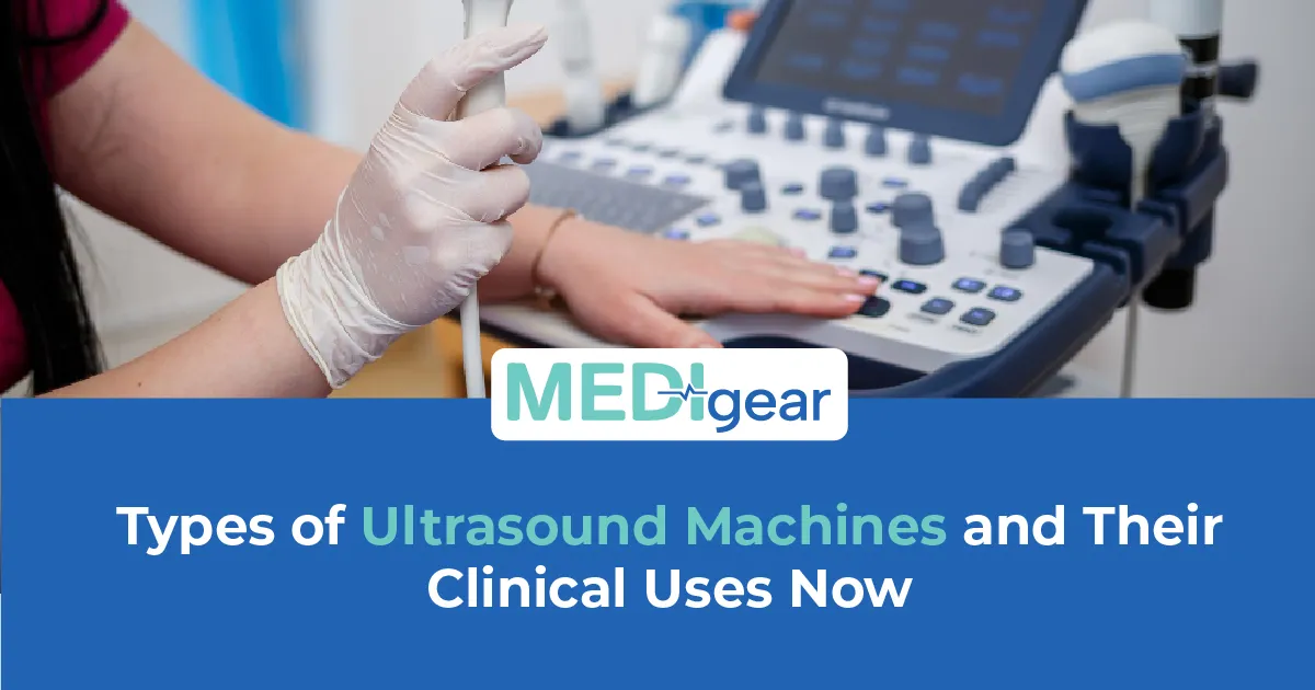
This guide covers types of ultrasound machines and clinical uses with the honest detail clinicians, owners, and procurement teams need. Medigear supplies certified ultrasound machines to hospitals, GP practices, and specialist clinics. Every point here comes from real clinical demand. Clinics sourcing these systems can explore the Medigear buyers portal for pricing, availability, and imaging procurement.
Cart-Based
Cart-based ultrasound machines are the workhorse of hospital imaging departments and large clinics. Large monitor. Keyboard. Multiple probe ports. Doppler. Elastography. Contrast. High quality. Deep. Fifty patients a day? Cart-based. Power. Storage. Quality. Workflow. Not portable. Wheels between rooms. ICU or community? Different type.
Portable
Portable ultrasound machines bring imaging to the patient. Wheeled portables fit through doors. Laptop-style on a trolley — reach the wards. Sacrifice depth and processing. Gain mobility. Ward assessment — effusion, cardiac, DVT. Portable at the bedside gives the answer that the scan booked next week cannot. Image at the bedside. Decision in the room. Not the radiology report. Ultrasound machine manufacturers wanting to list portable, cart-based, and point-of-care systems where hospitals and clinics are searching can reach buyers through the Medigear advertising platform.
POCUS
Point-of-care ultrasound — POCUS — is the fastest-growing clinical use. Handheld or compact. Treating clinician. At the moment of need. Emergency medicine. Intensive care. GP. Anaesthesia. One question at a time. Free fluid? Heart contracting? Bladder full? Which side? Does not replace the diagnostic scan. Answers the question now. Change management now. Small device. Specific training. Large clinical impact. Reach out to our team for guidance on matching ultrasound machine type to your clinical setting and patient volume.
Transducers
Transducers determine what each ultrasound machine can see. Curvilinear — low frequency, wide. Penetrates deep. Abdomen. Obstetrics. Pelvis. Linear — high frequency, narrow. Superficial. Tendons. Nerves. Thyroid. Vessels. Phased array — small, deep. Between ribs. Cardiac. Lung. Endocavitary — transvaginal and transrectal. Close to the pelvis and prostate. The same machine body may take multiple transducers. Buy one — limit to one clinical use.
Doppler
Doppler modes add flow information to the image. Colour Doppler — direction. Red toward. Blue away. Power Doppler — flow without direction. More sensitive in low-flow. Spectral Doppler — pulsed and continuous wave. Measures flow velocity. Used in vascular work. Cardiac output. Renal stenosis. DVT. Fetal wellbeing. No Doppler capability — cannot answer flow questions.
3D and 4D
3D and 4D ultrasound add volume to the image. 3D gives a static volume. 4D adds real-time motion. Obstetrics — fetal anatomy, face, cardiac. Gynaecology — uterus and ovary. Not every unit needs it. Fetal medicine — yes. Vascular lab — no. Our guide to Creutzfeldt-Jakob disease covers the diagnostic monitoring tools used in neurological conditions — the same principle applies when choosing ultrasound machine capability. Match the tool to the clinical question. Not the other way.
CEUS
Contrast-enhanced ultrasound — CEUS — uses microbubble contrast agents to assess organ perfusion and characterise lesions. Liver. Kidney. Spleen. Indeterminate on B-mode — characterised with contrast. The machine must support the contrast software. Not all do. Specialist. Not general-purpose. Our guide to essential eye screening devices covers the importance of matching diagnostic tools to clinical specialities — the same discipline applies when a team asks whether they need CEUS capability or whether a standard B-mode machine answers all the questions their patient population presents.
Elastography
Elastography measures tissue stiffness. Tissue stiffer than normal — fibrotic, malignant, or inflamed. Liver fibrosis assessment. Breast and thyroid lesion characterisation. The shear wave gives a quantitative stiffness value. Strain gives a relative comparison. Hepatology. Breast imaging. Thyroid. Not an emergency. Not obstetrics. The Hepatology unit needs it. Delivery suite does not.
Pre-Purchase Definition
Can your clinic define, before purchase, which clinical questions the ultrasound machine must answer — and which transducers those questions require? Procedures. Transducers. Software modes. Before the budget. Not after the machine arrives and the first case shows what is missing. Suppliers of cart-based, portable, and point-of-care ultrasound machines can register through the Medigear supplier portal to connect with hospitals and clinics defining their imaging requirements.
Training
Does your team have a structured training programme for every clinician who will operate the ultrasound machine? Image acquisition is a skill. Probe position. Depth. Gain. Doppler angle. The image that looks right and the image that is right are not always the same. Train to competency. Not familiarity. Protects from the confident misdiagnosis. Companies seeking long-term collaboration on ultrasound machine supply, servicing, and clinical training packages can explore the Medigear partnership programme for ongoing opportunities beyond a single order.
Portable vs POCUS
Does your team know the difference between a portable ultrasound machine and a point-of-care ultrasound device — and use each for the right task? Portable means the machine travels to the patient. POCUS means the clinician uses it at the bedside to answer one question. Both are mobile. One is a diagnostic imaging tool. One is a clinical decision aid. Using a portable as a POCUS device works. Using a POCUS device as a full imaging tool does not. Know the difference. Use accordingly.
Buyer Responsibility
The ultrasound machine does not choose its clinical setting. The buyer does. Get it right before the machine arrives — not after the first case shows what is missing.
Scope Creep
Does your department track which clinical questions each ultrasound machine type is being used to answer — and whether any are being used outside their intended scope? POCUS is used for a full abdominal scan. It was not built for general-purpose fetal cardiac use. Portable for contrast it cannot perform. Matching use to type is not paperwork. It keeps imaging accurate and defensible.
Transducer Bundle
Can your procurement team negotiate a transducer bundle — machine body plus multiple probes — that covers the full clinical scope from day one? One probe at purchase, three more later — costs more than the bundle agreed upfront. Define probes before purchase. Negotiate the bundle. Full capability from day one.
Service Evolution
What does your clinic do when a new clinical service is added that requires an ultrasound mode the current machine does not support? Elastography for hepatology. CEUS for oncology. Right for last year's service. It may not be right for next year. Review imaging capability against the clinical service. Keep them aligned.
Incident Log
Does your imaging team log every probe-related incident — dropped transducer, cable damage, patient complaint about probe temperature or pressure? Incident log captures every issue. Pattern found before it becomes the claim. Dropped twice in a month — protocol change. Cable fraying — supplier conversation. Log proves the team manages equipment. Not just patients.
End of Life
How does your team handle the ultrasound machine at the end of its useful life? Planned replacement. Not an emergency replacement when the machine fails mid-list and the next available slot is four weeks away. Capital planning builds replacement into the cycle before the machine fails. Budget before the failure funds the next machine. No budget — delay.
Maintenance
Does your clinic have a plan for ultrasound machine maintenance — annual service, transducer inspection, and software updates? Fraying cable — artefact. Cracked probe — infection risk. Software not updated may lack the modes the current protocol needs. Annual service. Log it. File it. Ready for inspection.
Image Consistency
Can your imaging team compare images over time using the same ultrasound machine settings? The liver assessed six months ago and the one today must be comparable. Same depth. Gain. Frequency. Position. Consistent acquisition drives the comparison. Machine quality alone is not the answer.
Machine Failure
What does your clinic do when the ultrasound machine fails during a diagnostic list? Service contract. Response time guaranteed. Loan machine during repair. Referral pathway. List stopped by a failed machine costs more than the contract that prevents it.
Probe Infection Control
Does your team know the cleaning and infection control requirements for every transducer in use? Endocavitary probes — high-level disinfection. Between every patient. Intact skin probes — intermediate. Wrong solution. Wrong level. Transducer transmits infection. Not optional. Regulated. Inspectable.
Why Choose Medigear
Medigear supplies certified cart-based, portable, and point-of-care ultrasound machines with a full range of transducers and imaging modes to hospitals, clinics, and specialist units across the UK. Whether you are equipping a new department, adding POCUS capability, or replacing an ageing system, our team matches the right ultrasound machine to your clinical questions and your budget. Reach out to our team for guidance built around the images your patients need — and the machines that produce them reliably every time.
Conclusion
What makes one ultrasound machine right for one job and wrong for another? The transducer. The software. The purpose it was built for. Small GP practice. Joint injections. Could not see the tendon sheath. One upgrade — portable, linear probe. Visible in real time. Hit rate up. Machine not wrong. Just wrong for that job. Cart-based for radiology. Portable for the ward. POCUS for the bedside question. Curvilinear for depth. Linear for tendons. Phased array for cardiac. Doppler for flow. Elastography for stiffness. CEUS for lesion characterisation. Define the questions before the budget. Define the transducers before the purchase. Train to competency. Not familiarity. Log every incident. Service every year. Plan every replacement. Medigear stands alongside imaging teams with certified ultrasound machines, transducers, and the honest support that clinical imaging demands. Speak to our team today — because the right ultrasound machine for the right clinical use must be chosen before the first patient, not after the first case reveals what is missing.
⚠️ This post is for general information only. We do not sell medications or provide prescriptions — Medigear.uk is a medical equipment supplier only.