Root canal treatment requires extreme precision to clean and seal tiny, complex canals inside a tooth. Even minor inaccuracies can leave behind infected tissue, leading to treatment failure or reinfection.
Dental operating microscopes have transformed the way endodontists perform root canal therapy—offering unparalleled magnification, illumination, and accuracy for better clinical outcomes.
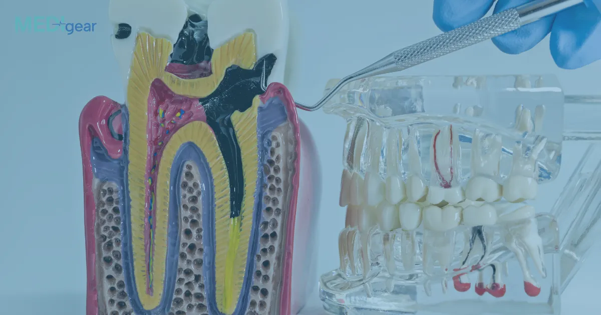
The Role of Microscopes in Modern Endodontics
A dental microscope is an advanced optical instrument designed to provide high magnification and enhanced lighting during dental procedures.
In root canal therapy, it helps clinicians visualize fine details of tooth anatomy that are invisible to the naked eye or traditional loupes.
By magnifying the operating field up to 25 times and illuminating deep structures with coaxial light, dental microscopes make the treatment more precise, efficient, and minimally invasive.
1. Enhanced Visualization of Canal Anatomy
Root canal systems can have complex variations, including multiple or curved canals.
With a dental microscope, dentists can detect accessory canals, narrow passages, and calcified areas that might otherwise go unnoticed.
This improves cleaning and shaping accuracy, reducing the risk of missed infections.
2. Improved Detection of Cracks and Fractures
Microscopic visualization allows clinicians to identify fine cracks or fractures in the tooth structure that are often invisible in standard exams.
Early detection helps determine whether a tooth can be saved or needs alternative treatment, ensuring the most conservative approach possible.
3. Increased Precision in Cleaning and Shaping
Microscopes enable endodontists to remove debris and infected tissue with exceptional precision.
Better visibility ensures complete debridement of root canals while preserving healthy tooth structure, which is vital for long-term tooth strength.
4. Greater Accuracy in Locating Canal Openings
Locating canal orifices—especially in molars—can be difficult. Dental microscopes guide the clinician in identifying these openings accurately, reducing procedural errors and unnecessary removal of dentin.
5. Enhanced Sealing and Filling Quality
The microscope provides a clear view of the canal during obturation (filling), ensuring complete sealing of the canal system.
This minimizes the chances of leakage or reinfection, significantly improving the success rate of root canal treatments.
6. Reduced Treatment Time and Complications
With enhanced visibility and precision, the dentist can perform procedures more efficiently, reducing the number of treatment sessions.
It also lowers the likelihood of complications such as perforations or missed canals.
7. Better Ergonomics and Documentation
Dental microscopes improve working posture for clinicians, minimizing fatigue during long procedures.
Many modern microscopes are equipped with digital imaging and recording systems, enabling real-time documentation and patient education.
Clinical Benefits for Patients
- Higher treatment success rates
- Less post-procedure discomfort
- Faster recovery
- Preservation of natural tooth structure
- Fewer repeat treatments
In short, dental microscopes enhance both the precision of care and the patient experience.
Final Thoughts
Dental microscopes have become indispensable in endodontics, setting a new standard for accuracy in root canal therapy.
By combining magnification, illumination, and digital visualization, they enable clinicians to perform treatments with greater confidence and precision—ensuring healthier, longer-lasting results for patients.
Disclaimer:
This article is for informational purposes only and does not replace professional dental advice. Always consult a qualified dentist or endodontist for proper diagnosis and treatment of dental conditions.