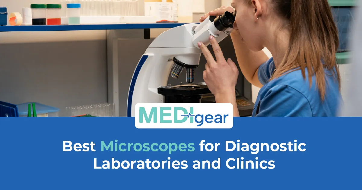
What if the diagnosis depended on one cell that the naked eye could never see? What if the difference between cancer and not cancer, malaria and not malaria, TB and not TB sat on a glass slide under a lens that was either sharp enough to show it or too cheap to resolve it? What if the microscope in the lab was the reason the report said normal when the tissue was not? That is what a bad unit does. It does not blur the image. It blurs the diagnosis. And in a diagnostic lab, a blurred diagnosis is a missed disease.
She ran a small pathology lab attached to a district hospital. The machine was twelve years old. Optics yellowed. Stage wobbled. Condenser stuck. Twenty minutes on a blood film that should have taken five. Not because the cells were complex. Because the image was too poor to trust. She reported what she could see. But she knew what she could not see was the problem. A new microscope — LED light, plan achromatic lenses, smooth stage — arrived on a Monday. By Friday, she had picked up a case of chronic lymphocytic leukaemia that the old microscope would have shown as normal lymphocytes. Optics changed. Diagnosis changed. Life changed. All from a lens.
This guide covers how to choose the best microscope for diagnostic laboratories and clinics with the honest detail that lab managers, pathologists, and procurement teams need. Medigear supplies certified microscopes to hospitals and labs across the UK — and every point here comes from real diagnostic demand, not catalogue specs.
Resolution
A microscope magnifies and resolves what the eye cannot see. But magnification without resolution is empty. Bigger blur. Still a blur. Resolution is the power to tell two points apart. It depends on the lens. The aperture. The condenser. The light. A diagnostic unit must resolve at the cell level. The gap between benign and malignant sits in fractions of a micron.
Brightfield
Brightfield microscopy is the foundation of diagnostic lab work. Stained tissue, blood films, urine sediment, and cytology all read under brightfield. Image quality depends on the lenses, the condenser, and the light. All working together. Koehler illumination — adjusting the condenser to produce even, glare-free light — is the baseline. Without Koehler, the image is too uneven for clinical reporting.
Objective Lenses
Objective lenses define the microscope. Plan achromatic lenses correct curvature and colour fringing. Flat. Sharp. Edge to edge. Semi-plan corrects only the centre. Non-plan curves, the edges — peripheral cells unreadable. For diagnostics, plan achromatic is the minimum. Plan apochromatic — all colour wavelengths corrected — gives the best image. Costs more. Match lens quality to the work. Blood films and histology need a plan achromatic. Fluorescence needs a plan-apochromatic.
Mechanical Stage
The stage moves the slide in precise steps — letting the user scan without missing areas. A stage that sticks, jumps, or drifts makes scanning unreliable. Smooth coaxial X-Y controls are the standard for any diagnostic unit. Playing on the stage turns a ten-minute scan into twenty minutes of frustration.
LED Illumination
LED illumination has replaced halogen in modern microscopes — and for good reason. LEDs last tens of thousands of hours. Halogen lasts for hundreds. Consistent colour. Less heat. Less power. No warm-up. And they do not yellow with age, keeping colour true across the life of the unit. For diagnostics, LED is not optional. First slide to last — colours stay true.
Linked Guides
For labs managing microscopes alongside broader diagnostic equipment, our guide to the best nebulisers covers the respiratory devices that depend on the lab results microscopes help produce — because a sputum culture read under the microscope guides the nebulised antibiotic the patient receives. Our guide to patient monitoring on a budget covers the bedside tools that work alongside lab diagnostics — because the blood film the microscope reads shapes the treatment the monitor tracks.
Binocular vs Trinocular
Binocular versus trinocular heads change what the microscope can do beyond visual reporting. Binoculars suit routine viewing. Trinocular adds a camera port. Digital capture. Remote review. Telepathology. Archiving. For labs building digital pathology, teaching, or QA systems, trinocular is the platform that grows with the service.
Digital Camera
Digital cameras attached to the microscope capture what the eye sees — and store it for review, comparison, audit, and teaching. A proper camera with measurement and annotation software gives the lab a permanent record of every key finding. A phone held to the eyepiece gives a blur that proves nothing. Invest in a proper camera if the images must stand up to scrutiny.
Fluorescence
Fluorescence microscopy uses UV or specific-wavelength light to excite fluorescent dyes bound to cellular targets. Essential for immunofluorescence. Detecting antibodies in the kidney, skin, and autoimmune cases. It needs excitation filters, barrier filters, and a dedicated fluorescence light source. Not every lab needs it. But every lab reporting immunofluorescence must have a unit that does it right.
Phase Contrast
Phase contrast microscopy makes transparent, unstained specimens visible by converting differences in light phase into differences in brightness. Used for urine sediment, unstained cells, and live observation. Phase-contrast microscopy adds what brightfield cannot — the ability to see structures that staining would alter or destroy.
Ergonomics
Ergonomics shape how long a microscopist can work before fatigue, neck pain, and eye strain force them to stop. Tilting heads. Adjustable height. Padded rests. Low controls. These cut the physical cost of hours at the eyepiece. A unit that forces hunching, reaching, or craning causes problems that pathologists carry for life. Buy ergonomic. The body behind the eyepiece is not replaceable the way the machine is.
Maintenance
Maintenance keeps the microscope accurate between services. Lenses need cleaning with proper tissue. The condenser needs realignment after lamp swaps. The stage needs lubricating. Oil must come off after every use. A unit not maintained drifts from the image it was bought to deliver — and the diagnosis drifts with it.
Certification
Certification matters. CE marking and IVD compliance confirm the microscope meets the standards a diagnostic lab demands. A teaching or hobbyist unit in a clinical setting may lack the optical quality and the cert that reporting requires. Buy IVD-certified. The report carries the weight of the microscope that produced it.
Backup
Can your lab run a second microscope when the primary fails mid-list? A single-unit lab stops reporting the moment the machine breaks. Slides wait. Results wait. Clinicians wait. One backup — even a simpler model — keeps the lab running while the primary is serviced. The cost of a spare is nothing against a day without diagnoses.
Training
Does your lab train every new user on Koehler alignment before they touch a slide? A microscope out of alignment produces an image that looks acceptable but is not diagnostic. Colours shift. Contrast drops. Edges blur. A trained user sets Koehler in under a minute. An untrained one works all day on an image that was never right. Training costs nothing. Bad alignment costs diagnoses.
Cost Per Year
How do you compare microscope costs across brands? Not by sticker price. By cost per diagnostic year. Add the purchase, lenses, camera, bulbs, maintenance, and expected lifespan. Divide by years of diagnostic use. A cheap microscope that needs new objectives in two years costs more per year than a quality one that runs ten years without a fault. Buy the unit that costs less per slide. Not less per invoice.
Teaching Heads
Multi-head teaching microscopes let a trainer and student view the same slide at the same time — essential for pathology training, competency sign-off, and second-opinion review. A lab without a teaching head relies on description alone. A lab with one shows exactly what the trainer sees — and the student learns from the real image, not a verbal sketch.
Dark Field
Dark field microscopy lights the specimen from the side, making transparent objects glow against a dark background. It suits spirochaete detection, crystal identification in joint fluid, and screening of unstained specimens. Not every lab needs dark field. But the one that does cannot swap it for brightfield. The organisms it reveals are invisible under standard light.
Oil Immersion
Oil immersion objectives — typically the hundred-times lens — deliver the highest resolution a light microscope can achieve. They require a drop of immersion oil between the lens and the slide to eliminate the air gap that reduces resolution. Oil immersion is essential for blood film differentials, TB smear reading, and malaria parasite identification. A diagnostic microscope without a high-quality oil-immersion objective is a machine missing the lens that matters most for the cases that matter most.
Real-World Testing
Does your procurement team test the microscope under real diagnostic conditions before buying? A demo with a clean, prepared slide in a bright showroom tells you nothing about how the machine performs on a poorly stained biopsy at 4 pm under fluorescent strip lights. Test it in your lab. With your slides. Under your conditions. The microscope that looks perfect in a demo may struggle where it matters — and the one that wins in the lab is the one your pathologists will trust.
Why Choose Medigear
Medigear supplies certified microscopes, cameras, accessories, and optical equipment to hospitals, pathology labs, and clinics across the UK — with clear pricing, honest guidance, and after-sales support built for the demands of daily diagnostic work. Whether you are equipping a new lab, upgrading an ageing microscope, or adding digital imaging, our team matches the right optics to your diagnostic need. Reach out to our team directly for guidance built around the slides your lab reads — and the diagnoses that depend on every lens.
Conclusion
What if the microscope in the lab was the reason the report said normal when the tissue was not? That is not a hypothetical. It is a reality in every lab where yellowed optics, wobbling stages, and stuck condensers turn diagnostic microscopy into guesswork. One new microscope. One Monday morning. One case of leukaemia caught that the old machine would have called normal lymphocytes. Optics changed. Diagnosis changed. Life changed. All from a lens. Medigear stands alongside pathology teams with certified microscopes and the honest support that diagnostic accuracy demands. Speak to our team today — because the slide under the lens carries a patient's future, and the microscope that reads it must be sharp enough to show the truth.
⚠️ This post is for general information only. We do not sell medications or provide prescriptions — Medigear.uk is a medical equipment supplier only.