 Aman Yadav
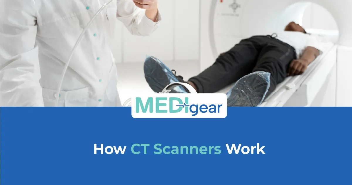
 Computed tomography has transformed diagnostic imaging by allowing healthcare professionals to examine internal body structures in far greater detail than standard X-rays. From emergency trauma assessment to cancer evaluation and surgical planning, CT scanners are now considered an essential part of modern medical imaging departments.
Unlike conventional imaging systems that produce a single flat image, CT scanners generate cross-sectional views of organs, bones, blood vessels, and soft tissues. This layered imaging capability helps clinicians detect abnormalities with improved clarity and speed.
Healthcare facilities evaluating imaging infrastructure often compare CT technology alongside broader radiology upgrades, especially when exploring how <a href= "https://medigear.uk/blog/how-digital-radiography-is-transforming-healthcare">digital radiography systems are reshaping diagnostic workflows</a>. Understanding how CT scanners operate also helps procurement teams make better equipment planning decisions.
Understanding the Core Principle Behind CT Imaging
A CT scanner combines rotating X-ray technology with advanced computer processing. During a scan, the machine captures multiple X-ray images from different angles around the patient's body. Powerful software then reconstructs those images into highly detailed cross-sectional slices.
Instead of viewing a single shadow-like image, clinicians can observe detailed internal structures layer by layer. This allows clearer visualisation of:
-
Bones and joints
-
Internal organs
-
Blood vessels
-
Tumours and lesions
-
Internal bleeding
-
Lung conditions
-
Spinal structures
The process is fast, non-invasive, and supports rapid clinical decision-making in emergency and routine diagnostic settings.
The Main Components Inside a CT Scanner
Modern CT systems integrate several technologies that work together simultaneously.
Rotating Gantry System – The large circular structure surrounding the patient houses the X-ray tube and detector array. It rotates rapidly during imaging to capture multiple projections from various angles.
Patient Positioning Table – The motorised table moves gradually through the gantry while scans are being performed. Precision movement is important for accurate image reconstruction.
X-Ray Generation Unit – This component produces controlled X-ray beams that pass through the body. Different tissues absorb radiation differently, creating the contrast needed for imaging.
Digital Detector Array – Detectors positioned opposite the X-ray source measure the radiation passing through the body and convert it into electrical signals.
Image Reconstruction Software – Advanced computing systems process raw scan data into detailed cross-sectional images that radiologists can interpret.
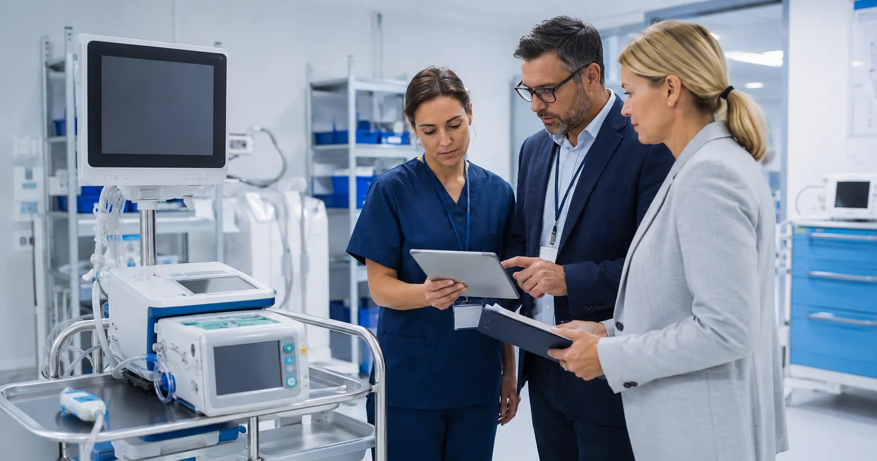
Hospitals expanding imaging services often coordinate CT investments with broader procurement planning strategies. Facilities comparing multiple technology suppliers may also explore verified sourcing options for healthcare equipment through Medigear. UK Buyers Network.
What Happens During a CT Scan?
The scanning process typically follows a structured workflow designed for speed and imaging accuracy.
Patient Preparation
Depending on the examination type, patients may be asked to:
-
Remove metallic objects
-
Change into hospital gowns
-
Avoid eating for several hours
-
Receive contrast material for enhanced imaging
Contrast agents are commonly used for vascular imaging and soft tissue visibility.
Image Acquisition
Once positioned on the scanning table, the patient moves through the gantry while the X-ray tube rotates continuously. Thin imaging slices are captured within seconds.
Modern systems can perform hundreds of image acquisitions during a single scan cycle.
Digital Image Reconstruction
The collected imaging data is processed using sophisticated algorithms. Radiologists then review the reconstructed images on specialised diagnostic workstations.
Many healthcare facilities that integrate advanced imaging also evaluate how portable systems improve workflow flexibility, particularly after reviewing <a href="https://medigear.uk/blog/portable-x-ray-machines-benefits-and-applications">portable imaging solutions used in emergency care environments</a>.
Why CT Scanners Produce Detailed Images
The strength of CT imaging lies in its ability to produce cross-sectional slices rather than overlapping shadow images.
Capturing Multiple Angles Simultaneously – The rotating gantry gathers imaging data from numerous positions around the body, improving anatomical detail.
Distinguishing Soft Tissue More Clearly – CT imaging offers far better soft tissue differentiation than conventional X-rays, especially in neurological, thoracic, and abdominal imaging.
Generating 3D Reconstructions – Advanced systems can reconstruct three-dimensional anatomical models for surgical planning and interventional procedures.
Supporting Rapid Emergency Assessment – Trauma departments rely heavily on CT technology because scans can identify life-threatening conditions within minutes.
Common Clinical Areas Where CT Imaging Is Used
CT scanners are used across multiple medical specialities and diagnostic workflows.
Emergency Trauma Evaluation – Rapid imaging helps detect fractures, bleeding, and internal injuries following accidents.
Neurological Imaging – Brain scans help identify strokes, haemorrhages, and other neurological abnormalities.
Cancer Detection and Monitoring – CT imaging supports tumour detection, treatment planning, and disease progression monitoring.
Cardiovascular: Assessment. Specialised CT protocols help visualise coronary arteries and vascular conditions.
Pulmonary Diagnostics – Chest CT scans provide detailed lung imaging for respiratory disease evaluation.
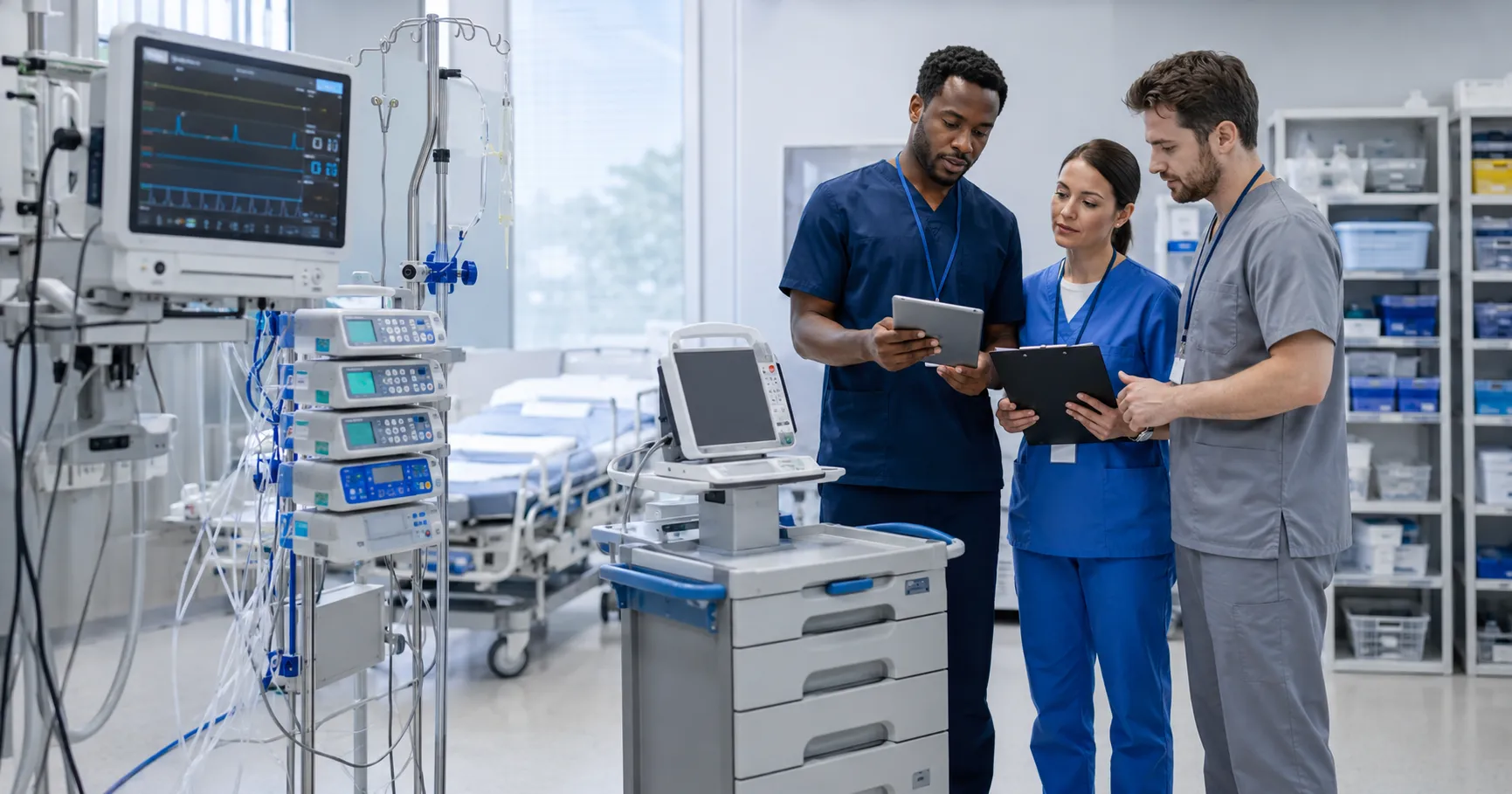
Facilities expanding advanced imaging capabilities frequently collaborate with certified imaging equipment suppliers through Medigear—UK Supplier Connections to evaluate installation requirements and long-term servicing support.
Different Types of CT Scanner Technologies
CT technology continues evolving to support faster imaging and improved diagnostic precision.
Slice-Based CT Systems
Older CT systems captured fewer slices per rotation, resulting in longer scan times. Modern multi-slice systems now generate significantly more image data during each rotation cycle.
Spiral and Helical CT Imaging
These scanners continuously rotate while the patient table moves steadily through the gantry. This produces faster image acquisition and smoother anatomical coverage.
Dual-Energy CT Technology
Dual-energy systems use two energy levels to improve tissue differentiation and material analysis.
AI-Assisted Reconstruction
Newer imaging software uses artificial intelligence to reduce noise, improve image clarity, and support lower radiation exposure.
Healthcare organisations researching future-ready imaging strategies may also participate in collaborative equipment initiatives through Medigear.UK PaUKership Programs.
Radiation Safety in CT Imaging
CT scanners use ionising radiation, making dose management an important consideration.
Modern systems include several technologies designed to reduce exposure while maintaining image quality.
Automated Dose Optimisation – Software adjusts radiation levels based on patient size and scan requirements.
Faster Scan Speeds – Reduced scanning time helps minimise unnecessary exposure.
Iterative Reconstruction Techniques – Advanced image processing allows lower-dose imaging without compromising diagnostic quality.
Healthcare facilities typically establish strict imaging protocols to maintain radiation safety standards.
Operational Factors Hospitals Consider Before Purchasing CT Systems
CT scanner procurement involves far more than selecting image quality specifications.
Installation Requirements – CT rooms require specialised shielding, cooling systems, and structural planning.
Service Accessibility – Reliable technical support and preventive maintenance are critical for minimising downtime.
Workflow Integration – Modern scanners must integrate with PACS, RIS, and hospital information systems.
Patient Throughput Capacity – High-volume hospitals prioritise systems that can handle continuous imaging demand.
Training and User Familiarity – Radiology staff training influences operational efficiency and image consistency.
Hospitals needing procurement guidance or installation coordination can reach the Medigear UK Support Team for operational assistance and sourcing information.
How CT Imaging Is Shaping Modern Healthcare
Diagnostic imaging continues shifting toward faster, more connected, and AI-enhanced systems.
Several trends are currently influencing CT technology adoption:
-
Low-dose imaging development
-
AI-assisted diagnostic support
-
Faster emergency imaging workflows
-
Cloud-based imaging integration
-
Advanced cardiac CT expansion
-
Portable CT deployment in critical care
As healthcare systems prioritise early diagnosis and operational efficiency, CT imaging remains central to modern clinical infrastructure planning.
Disclaimer
Medigear.uk is a medical equipment supplier and distributor. We do not provide medical advice, diagnosis, or treatment recommendations. All information is for educational and product awareness purposes only. Healthcare decisions should always be made by qualified medical professionals.