Colorectal Cancer Screening and Surveillance
The Olympus CF-H185I is widely deployed in national and opportunistic colorectal cancer screening programmes, enabling high-definition mucosal inspection, adenoma detection, and polypectomy during routine diagnostic colonoscopy. HDTV imaging combined with NBI supports optical diagnosis, DISCARD strategy adoption, and confident characterisation of diminutive polyps, improving adenoma detection rates in screening and post-polypectomy surveillance cohorts across hospitals and endoscopy centres.
Polypectomy and Endoscopic Mucosal Resection
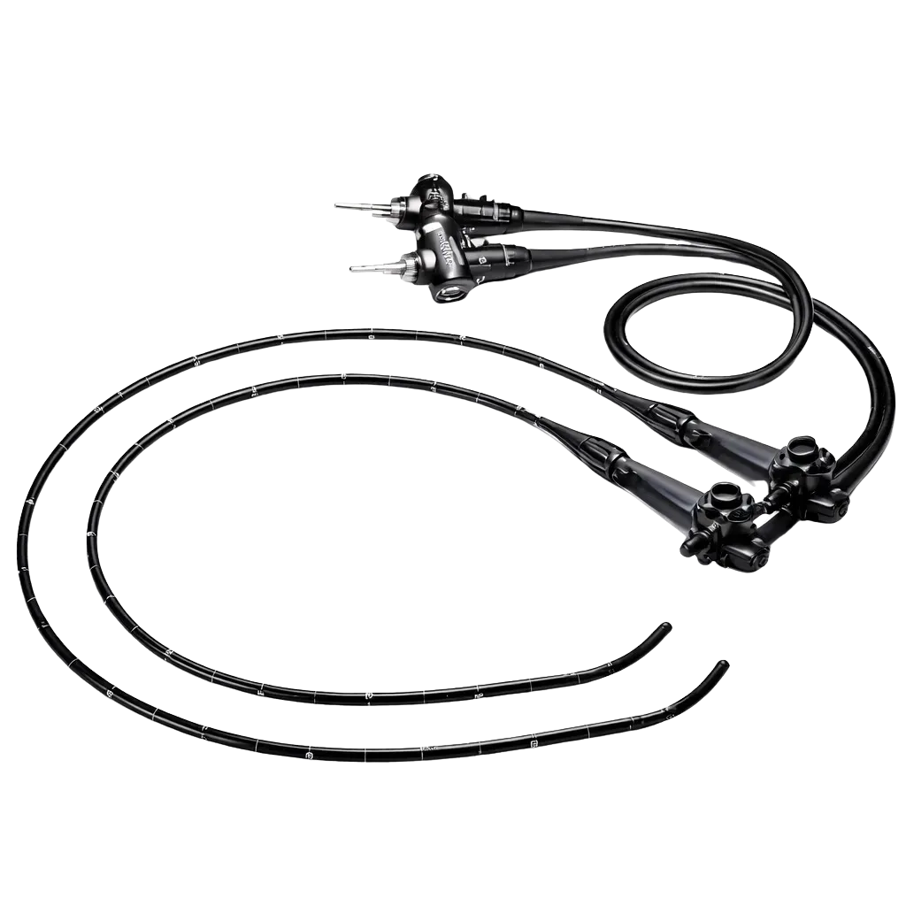
The 3.7 mm working channel accepts snares, hot biopsy forceps, clips, and EMR accessories for effective removal of sessile, pedunculated, and flat polyps. Variable Stiffness and precise angulation support therapeutic access to lesions in the caecum, flexures, and rectum, making the CF-H185I suitable for outpatient endoscopy units and tertiary therapeutic endoscopy services.
Inflammatory Bowel Disease Assessment
Gastroenterologists use the CF-H185I for ulcerative colitis and Crohn's disease evaluation, including disease-activity scoring, dysplasia surveillance, and targeted biopsy. HDTV imaging and NBI support accurate mucosal assessment and chromoendoscopy-style evaluation without dye, improving IBD diagnostic yield in hospital and specialist clinic settings.
Haemorrhage Investigation and Therapeutic Haemostasis
The CF-H185I supports urgent colonoscopy for lower gastrointestinal bleeding assessment, allowing identification of bleeding sources and endoscopic haemostasis through clipping, thermal coagulation, or injection therapy. The waterproof connector and rapid setup support same-day emergency workflows in hospital endoscopy units.
Diagnostic Biopsy and Targeted Tissue Sampling
Routine diagnostic colonoscopy with targeted biopsy for suspected colitis, ischaemia, or malignancy is supported by the 3.7 mm channel and precise angulation control. Community endoscopy units, hospital gastroenterology departments, and specialist colorectal clinics benefit from reliable tissue sampling and confident histological diagnosis.