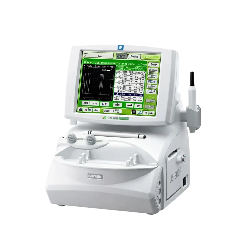
50MHz High-Frequency Ultrasound Imaging
The US-500P uses 50MHz high-frequency ultrasound to produce real-time cross-sectional imaging of the anterior segment with resolution not achievable by conventional ophthalmic ultrasonography. This frequency provides the detail required for accurate iridocorneal angle assessment, ciliary body imaging, and anterior uveal pathology characterisation.
Iridocorneal Angle Assessment
Real-time cross-sectional imaging of the iridocorneal angle enables direct measurement of angle width, identification of angle closure mechanisms, detection of plateau iris and peripheral anterior synechiae, and assessment of ciliary body rotation in complex glaucoma evaluation and surgical planning.
Ciliary Body and Posterior Iris Imaging
The US-500P visualises structures not accessible by slit lamp or OCT, including the ciliary body, posterior iris, zonules, and pars plana. This imaging supports assessment of ciliary body cysts, anterior segment tumours, phakic IOL clearance, and post-surgical anatomy evaluation.
Real-Time Imaging with Freeze and Image Capture
Real-time dynamic imaging with freeze-frame and image capture capability supports detailed review of anterior segment findings, serial comparison, and structured clinical documentation for patient records, specialist referral, and clinical audit.
Quantitative Angle Measurement
The US-500P supports quantitative measurement of anterior chamber angle parameters, providing numerical data on angle width and dimensions that support serial comparison and evidence-based glaucoma management decisions.
CE Certified for Ophthalmology Clinical Use
The US-500P carries CE marking in compliance with applicable medical device directives, satisfying procurement compliance for hospitals, specialist glaucoma units, and regulated ophthalmology settings. Full documentation is available from Medigear.uk.