DirectRay Amorphous Selenium Detector
The DirectRay digital image receiver uses amorphous selenium (a-Se) to convert X-rays directly into electronic signals, eliminating light conversion and bypassing image-degrading scattering. Direct conversion preserves image integrity with unrivalled sharpness and detail.
Industry's Largest 24×29 cm Field of View
The 24×29 cm digital detector — the industry's largest single-detector field of view — accommodates almost all breast sizes in a single exposure. The expanded field eliminates additional exposures for larger patients, supporting workflow efficiency across mammography services.
FAST Self-Adjusting Tilt Paddle
The FAST (Fully-Automatic Self-Adjusting Tilt) paddle automatically adapts to the breast's natural contour, providing uniform compression across the entire breast. The adaptive paddle improves image quality while minimising patient discomfort across screening and diagnostic services.
HTC High Transmission Cellular Grid
The HTC (High Transmission Cellular) anti-scatter grid significantly reduces radiation scatter and produces higher contrast images. The grid technology supports detailed lesion characterisation, microcalcification visualisation, and consistent image quality across diverse breast morphology.
Molybdenum Rotating Anode Tube
The molybdenum rotating anode X-ray tube features focal spots of 0.1 mm (small) and 0.3 mm (large), 300 HU heat capacity, and 60 HU/min heat dissipation rate. Reliable Mo tube performance supports consistent throughput and detailed acquisition across busy services.
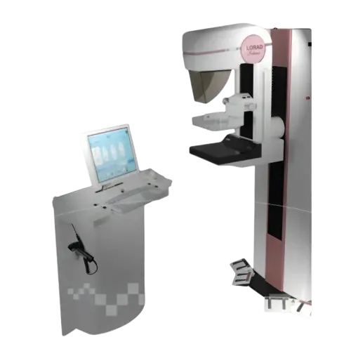
Acquisition Station with Integrated Control
The acquisition station features integrated X-ray control, intuitive image acquisition console, local storage and archiving, and trackball or function-key generator operation. Automatic exposure control with auto positioning and collimation supports streamlined daily mammography workflow.
DICOM Connectivity and Biopsy Support
DICOM 3.0 connectivity, ImageChecker CAD support, MIMS Mammography Image Management Solution, StereoLoc II breast biopsy device compatibility, and Peripheral Contrast Enhancement (PCE) support comprehensive workflow integration across screening, diagnostic, and biopsy applications.