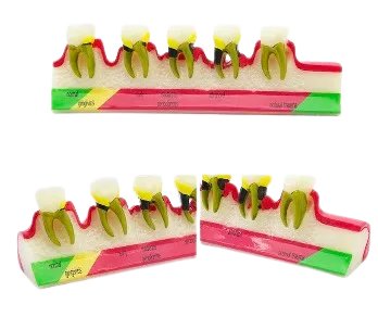
Colour-Coded Classification Zones
The model splits the arch into coloured zones, with each zone standing for a different state of gum health. This makes the move from one stage to the next easy to follow.
Healthy to Advanced Progression
One end shows healthy gums while the far end shows more advanced disease. Lining the stages up in order tells the story of how gum disease builds over time. It helps an audience grasp cause and effect at a glance.
Exposed Root and Support View
Part of the block is shown in section so the roots and the tissue around them are visible. As disease grows, the loss of support near the roots can be pointed out clearly.
Connected Row for Comparison
Because the teeth sit together on one block, healthy and diseased areas can be compared straight away. There is no need to switch between separate models. The side-by-side layout makes differences stand out.
Stable Display Base
The teeth and gum rest on a firm base that keeps the model steady on any surface. It stays in place while a clinician points to each zone. This suits both a busy teaching room and a quiet chairside talk.
Clear Visual Contrast
The colours and the exposed roots make the parts easy to tell apart at a glance. A patient or student can follow along with little set up. This keeps a short talk moving and holds attention.
Repeatable Demonstration Use
The model is made to be shown again and again during lessons or patient visits. It gives the same clear view each time it is used.