Enlarged Six-Times Scale
The model is shown at around six times life size, so fine detail becomes easy to see. Tiny features that are hard to point out in a real mouth stand out clearly here.
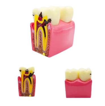
Healthy and Decayed Sides
One side of the model stays sound while the other shows decay setting in. Placing the two together lets a viewer compare them at a glance. The contrast makes the effect of caries plain without a long explanation.
Sectioned Inner View
The tooth is opened in section to reveal the enamel, the dentine, the pulp, and the canals. A dentist can trace how decay moves from the surface toward the nerve. This turns a hidden process into something easy to see.
Clear Decay Markings
The decayed areas are shown in dark tones that stand out against the rest of the tooth. This makes the path of the damage simple to follow.
Coloured Inner Parts
The pulp and canals are picked out in colour so the inside of the tooth is easy to read. This helps a viewer link the decay to the parts it threatens. It keeps an explanation clear and well ordered.
Stable Gum Block
The tooth sits in a firm gum block that keeps the model steady on any surface. It stays in place while a dentist points to each part. This suits both a teaching room and a quiet chairside chat.
Repeatable Demonstration Use
The model is made to be shown again and again across many appointments and lessons. It gives the same clear view each time it is used.