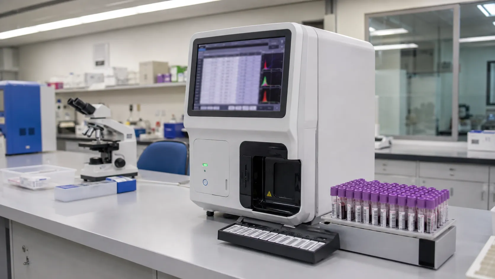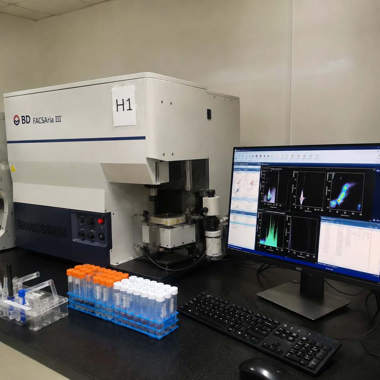
Flow cytometry is a laboratory technique that measures multiple physical and chemical characteristics of single cells as they pass in a fluid stream past laser beams, allowing identification and quantification of distinct cell populations in a suspension. Cells are stained with fluorescently labelled antibodies, streamed one by one past the lasers, and the scattered light and emitted fluorescence from each cell are recorded, so thousands of cells per second are characterised individually.
This guide is for laboratory managers and biomedical scientists specifying flow cytometers for immunophenotyping, research or clinical diagnostics. It explains how fluorophores report on cells, the difference between an analyser and a cell sorter, and the configuration, standards and running costs a buyer should weigh before purchase.
How Flow Cytometry Reads Single Cells
Hydrodynamic focusing is the core trick: the sample is injected into a faster-moving sheath fluid that narrows the stream so ce. Hence, line up single file through the laser interrogation point. As each cell passes, forward scatter reports roughly on size and side scatter on internal granularity and complexity. At the same time, und fluorophores absorb the laser light and re-emit at longer wavelengths. Optical filters and detectors separate these emissions into channels, and the instrument records a set of parameters for every event, building a multi-dimensional picture of the population.
Fluorophores and Immunophenotyping
The power of flow cytometry comes from fluorophore-conjugated antibodies that bind specific surface or intracellular markers. Because each fluorophore emits in a characteristic wavelength band, several can be used together, so a single tube reports on many markers at once. Immunophenotyping uses this to define cell types by their marker profile, for example identifying lymphocyte subsets by their cluster-of-differentiation markers. The number of colours an instrument can resolve, driven by how many lasers and detectors it carries, sets how deeply a laboratory can characterise a sample. More colours demand careful panel design and compensation or spectral unmixing to correct for the spectral overlap between dyes.
Analysers Versus Cell Sorters
Two instrument classes share the same optics but differ in what happens after measurement. An analyser measures cells and then sends them to waste; it answers how many cells of each type are present and what they express. A cell sorter adds the ability toto recover chosen populations physically. The most common approach breaks the stream into charged droplets, each ideally containing one cell; based on the real-time measurement, the instrument applies a charge and deflection fields steer droplets carrying target cells into collection tubes. Sorting lets a laboratory purify a population for culture, sequencing or functional assays, but it adds complexity, biosafety considerations for aerosols, and cost. Choose a sorter only where recovering live, defined cells is genuinely required; otherwise an analyser is simpler and cheaper to run.
Gating and Data Analysis
Raw events become populations through gating. On scatter and fluorescence plots, the operator or an automated routine draws boundaries that isolate the cells of interest and exclude debris, dodoublets nd dead cells. A consistent strategy is what makes results reproducible, so protocol-driven analysis, saved templates, and, in clinical work, validated software matter. Buyers should assess the acquisition and analysis software as seriously as the hardware, including how it handles compensation, batch analysis and export to the laboratory record.
Configuration a Buyer Must Specify
Flow cytometers are highly configurable, and the right build depends on the panels the laboratory runs. Key choices are the number and wavelengths of lasers, the number of fluorescence detectors and their filter sets, and whether the system uses conventional filter-based detection or spectral detection that captures the full emission spectrum for unmixing. Also weigh sample throughput, plate-based autosampling for high-volume immunophenotyping, and the volume and concentration handling for your specimen types. Over-specifying colours you will never stain wastes money; under-specifying forces compromises in panel design, so match the configuration to the assays on the menu now and realistically next.
Standards, Regulation and Quality
Where flow cytometry is used for clinical diagnosis, the laboratory works to ISO 15189 and validates each assay, with internal quality control and external quality assessment to keep results comparable. Clinical instruments, reagents and antibody panels are in-vitro diagnostic medical devices carrying UKCA or CE marking and regulated by the MHRA; the in-vitro diagnostic regulation (IVDR) affects which reagents are available as IVD-labelled versus research-use-only, which matters for a diagnostic service. Distinguish clearly at procurement between research-use and IVD-labelled reagents, because using research-only reagents in patient testing carries validation and governance obligations set out in UK guidance.
Reagents, Consumables and Running Costs
The recurring spend is dominated by fluorophore-conjugated antibodies, which are expensive and panel-specific, along with sheath fluid, calibration and setup beads, and cleaning solutions. Sorters add collection consumables and higher biosafety costs. When costing a platform, include daily quality-control beads that verify laser alignment and detector performance, the antibody cost per test across your panels, and waste handling for stained biological material. A realistic cost-per-result contract life, including these consumables, tells you far more than the capital price.
Procurement Checklist
-
Confirm UKCA or CE marking, MHRA registration and whether reagents are IVD-labelled or research-use under IVDR.
-
Match laser count, wavelengths and detector channels to your fluorophore panels.
-
Decide analyser versus sort, and whether live cells are genuinely needed.
-
Assess acquisition and analysis software, compensation or spectral unmixing and audit trails.
-
Check throughput, plate autosampling and specimen-volume handling for your workload.
-
Cost antibodies, setup beads, sheath and cleaning fluids per reportable result.
-
Confirm LIS or data-system connectivity and secure export of patient results.
-
Agree service cover, laser and fluidics spares, and application-support access.
Service, LIS Connectivity and Support
Lasers, detectors and fluidics need periodic service and occasional replacement, so compare preventive-maintenance schedules, response times and the cost of major parts, and ask whether a loan instrument is offered during a long repair. For diagnostic use, confirm how results and quality-control data connect to the LIS or a validated data system, since manual transcription is both slow and error-prone. Application support is especially valuable in flow cytometry, where panel design, compensation and troubleshooting spectral overlap draw on specialist expertise that a good vendor provides alongside the hardware.
Sample Preparation, Controls and Setup
A flow cytometry result depends heavily on sample preparation; blood immunophenotyping usually involves staining with the antibody panel and then lysing red cells, while other specimens need careful single-cell suspension so debris and clumps do not confound the read. Viability staining separates dead cells that bind antibody non-specifically, and doublet discrimination using scatter pulse geometry removes stuck-together cells that would otherwise appear as false populations. Getting these steps consistent is what makes gating reproducible from day to day.
Instrument setup adds its own controls. Daily quality-control beads verify laser alignment, detector sensitivity and fluorescence stability, and target values track performance over time. For multicolour panels, compensation controls and single-stained references for each fluorophore let the software correct spectral spillover, while spectral systems use reference spectra for unmixing instead. Buyers should confirm the platform makes this setup efficient, because a cytometer that is slow or fiddly to standardise each morning erodes the productivity its optics promise.
Conclusion
Flow cytometry identifies and quantifies single cells by the light they scatter and the fluorophores they carry, and a cell sorter goes further by physically recovering chosen populations. Specify the laser and detector configuration around the panels you actually run, decide honestly whether you need sorting, and cost the antibodies and service that dominate ownership. To compare cytometry platforms and consumables or arrange a laboratory quote, contact MediGear or set up a buyer account.
Disclaimer
This article is for informational purposes only. It is pu (medigear.uk) for genepublishes it ral information and procurement guidance, and is; itclinical, diagnostic, treatment, technical, engineering, legal or regu,latory advice, nor a product endorsement, guarantee or subs,titute for professional assessment. MediGear does not provide medical consultations. Buyers should consult their clinical, biomedical, estates and regulatory contacts, and the manufacturer's documentation, and independently verify all specifications, certifications, compatibility and suitability before purchase. Specifications, certifications and availability are correct at the time of publication and may change without notice. MediGear is a medical-equipment distributor and does not sell medicines or pharmaceutical products.