Introduction
Medical imaging has transformed the way doctors diagnose, monitor, and treat diseases. Among the most widely used imaging technologies are MRI, CT, and PET scans. While each plays a vital role in modern healthcare, they are designed for different purposes and rely on distinct technologies. Understanding their differences helps patients, caregivers, and even healthcare professionals make informed choices about diagnostic procedures.
MRI (Magnetic Resonance Imaging)
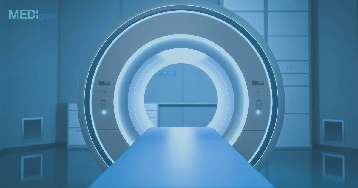
MRI uses powerful magnets and radio waves to generate detailed images of organs and tissues inside the body. Unlike X-rays or CT scans, MRI does not use ionizing radiation, making it safer for repeated use.
- How it works: MRI aligns hydrogen atoms in the body using a strong magnetic field. Radiofrequency pulses then disturb this alignment, and the returning signals are captured to form images.
- Best suited for: Soft tissues such as the brain, spinal cord, muscles, ligaments, and internal organs.
- Advantages: Provides highly detailed images, especially for neurological, musculoskeletal, and cardiovascular conditions. It’s also non-invasive and radiation-free.
- Limitations: More expensive and time-consuming compared to CT. Patients with pacemakers or certain implants may not be able to undergo MRI.
CT Scan (Computed Tomography)
CT scans use X-ray technology combined with computer processing to create cross-sectional images of the body. Unlike regular X-rays, CT provides detailed 3D images, giving doctors a clearer view of bones, blood vessels, and internal organs.
- How it works: The CT machine rotates around the patient, taking multiple X-ray images from different angles. A computer then processes these into detailed slices of the body.
- Best suited for: Detecting fractures, tumors, internal bleeding, lung and chest conditions, and emergency injuries.
- Advantages: Quick and widely available, making it highly useful in emergency medicine. Provides excellent detail for bone and internal injuries.
- Limitations: Uses ionizing radiation, which may not be suitable for repeated scans. Offers less soft-tissue detail compared to MRI.
PET Scan (Positron Emission Tomography)
PET scans focus not just on anatomy but on metabolic and biochemical activity. By using a small dose of radioactive tracer, PET scans reveal how organs and tissues are functioning at the cellular level.
- How it works: A radioactive tracer (often glucose-based) is injected into the patient. Cancer cells and other high-activity tissues absorb more of the tracer. The PET scanner detects the radiation emitted and creates images of metabolic activity.
- Best suited for: Cancer detection and monitoring, assessing brain disorders like Alzheimer’s disease, and evaluating heart function.
- Advantages: Provides functional information, showing how organs and tissues are working, not just how they look. Especially powerful when combined with CT or MRI for both structural and functional detail.
- Limitations: Involves exposure to radioactive tracers. More expensive and less widely available compared to CT or MRI.
Key Differences in Use
- MRI is ideal for soft tissue imaging and neurological conditions.
- CT scans excel in emergencies, trauma care, and bone imaging.
- PET scans reveal functional activity, making them indispensable in cancer detection and treatment monitoring.
Each technology complements the others rather than replacing them. In fact, many hospitals now use hybrid machines such as PET-CT or PET-MRI to combine functional and structural imaging for more accurate diagnosis.
Conclusion
MRI, CT, and PET scan machines each serve distinct purposes in medical imaging. MRI offers unparalleled detail for soft tissues, CT scans provide rapid and clear images of internal structures, and PET scans reveal how organs are functioning at the cellular level. Together, they form the backbone of modern diagnostics, helping physicians deliver precise and effective care.


