Magnetic Resonance Imaging (MRI) has become one of the most widely used imaging techniques in modern medicine, offering clear and detailed views of internal body structures without the use of radiation. A related technology, functional Magnetic Resonance Imaging (fMRI), extends the capabilities of MRI by mapping brain activity in real time. While the two modalities share a common foundation, their purposes and applications differ significantly.
What Is MRI?
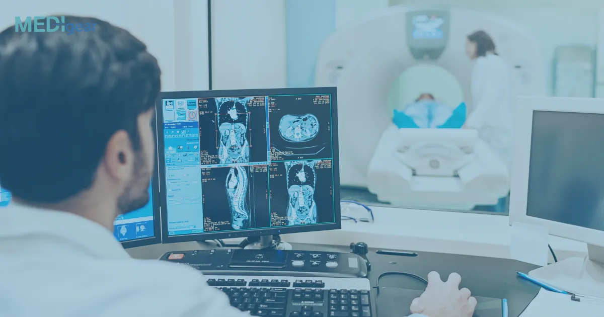
MRI is a non-invasive imaging technique that uses strong magnetic fields and radio waves to create detailed images of organs, tissues, and skeletal structures. It is especially useful for soft tissue imaging, making it invaluable in diagnosing conditions of the brain, spinal cord, joints, and internal organs.
Key uses of MRI include:
- Detecting tumors, infections, and abnormalities in the brain and spinal cord
- Imaging joints, ligaments, and cartilage for musculoskeletal injuries
- Evaluating heart structures and blood vessels
- Diagnosing conditions in the liver, kidneys, and other abdominal organs
What Is fMRI?
Functional MRI (fMRI) is a specialized form of MRI that measures and maps brain activity. Instead of focusing only on structure, fMRI detects changes in blood oxygen levels (known as the BOLD signal – Blood Oxygen Level Dependent), which increase in regions of the brain that are more active.
Key uses of fMRI include:
- Mapping brain regions responsible for movement, speech, and sensory processing
- Supporting neurosurgical planning by identifying critical brain areas
- Studying brain function in neurological and psychiatric conditions
- Advancing research in cognitive science and psychology
MRI vs. fMRI: Key Differences
|
Aspect |
MRI |
fMRI |
|
Purpose |
Produces detailed anatomical images |
Maps brain activity based on blood flow |
|
Focus |
Structural imaging |
Functional imaging |
|
Applications |
Diagnosis of physical conditions (tumors, injuries, organ diseases) |
Neuroscience, brain mapping, surgical planning |
|
Output |
Static images of body structures |
Dynamic images showing brain activity patterns |
|
Duration |
20–60 minutes depending on the scan |
Similar duration, but may involve tasks for brain activation |
Conclusion
MRI and fMRI share the same technological foundation but serve very different purposes. MRI provides detailed structural images of the body, essential for diagnosing diseases and injuries, while fMRI focuses on functional activity in the brain, playing a key role in research and neurosurgery. Together, these imaging tools broaden the understanding of both anatomy and function, supporting advances in healthcare and neuroscience.
Disclaimer: This article is for informational purposes only and does not constitute medical advice. For diagnosis, treatment, or medical concerns, always consult a qualified healthcare professional.